WB
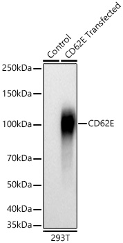
Western blot analysis of lysates from wild type (WT) and 293T cells transfected with CD62E using CD62E Rabbit mAb at 1:1000 dilution incubated overnight at 4℃. Secondary antibody: HRP-conjugated Goat anti-Rabbit IgG (H+L) at 1:10000 dilution. Lysates/proteins: 20 μg per lane. Blocking buffer: 3% nonfat dry milk in TBST. Detection: ECL Basic Kit.Exposure time: 30s.ICC/IF
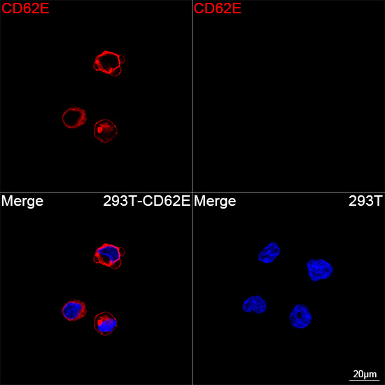
Confocal imaging of 293T cells transfected with CD62E using CD62E Rabbit mAb (dilution 1:200) followed by a further incubation with Cy3 Goat Anti-Rabbit IgG (H+L) (dilution 1:500) (Red). DAPI was used for nuclear staining (Blue). Objective: 100x.FC
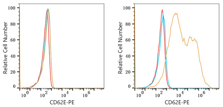
Flow cytometry: 1X10^6 CHO cells (negative control,left) and CHO (Transfection,right) cells were surface-stained with CD62E Rabbit mAb (2 μg/mL,orange line) or Rabbit IgG isotype control (2 μg/mL,blue line), followed by PE Goat anti-Rabbit pAb staining. Non-fluorescently stained cells were used as blank control (red line).FC
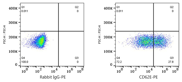
Flow cytometry: 1X10^6 CHO (Transfection) cells were surface-stained with Rabbit IgG isotype control (2 μg/mL,left) or CD62E Rabbit mAb (2 μg/mL,right), followed by PE Goat anti-Rabbit pAb staining.| Product Name | CD62E Rabbit mAb |
|---|---|
| Antibody Type | Primary Antibodies |
| Immunogen | Recombinant protein. |
| Clonality | Monoclonal |
|---|---|
| Isotype | IgG |
| Host Species | Rabbit |
| Tested Applications | FCICC/IFWB |
| WB:1:500-1:1000 ICC/IF:1:50-1:200 FC:1:500-1:1000 |
|
| Species Reactivity | Mouse |
| Concentration | 1mg/ml |
| Purification | Affinity purified |
| Gene Symbol | Sele |
|---|---|
| Gene Synonyms | Elam CD62E ELAM-1 LECAM2 E-selectin |
| Gene Full Name | selectin, endothelial cell |
| Gene Summary | Predicted to enable several functions, including oligosaccharide binding activity; phospholipase binding activity; and sialic acid binding activity. Involved in positive regulation of leukocyte tethering or rolling. Acts upstream of or within positive regulation of leukocyte migration. Predicted to be located in several cellular components, including caveola; clathrin-coated pit; and perinuclear region of cytoplasm. Predicted to be active in external side of plasma membrane and extracellular space. Is expressed in several structures, including adrenal gland; genitourinary system; incisor; integumental system; and limb segment. Human ortholog(s) of this gene implicated in IgA glomerulonephritis; brain ischemia; cerebrovascular disease; and coronary artery disease. Orthologous to human SELE (selectin E). [provided by Alliance of Genome Resources, Jul 2025] |
| Alternative Names | Elam CD62E ELAM-1 LECAM2 E-selectin |
| Molecular Weight(MW) | 66kDa(Observed MW 115kDa) |
| Cellular Localization | Cell membrane, Single-pass type I membrane protein. |
WB
Western blot analysis of lysates from wild type (WT) and 293T cells transfected with CD62E using CD62E Rabbit mAb at 1:1000 dilution incubated overnight at 4℃. Secondary antibody: HRP-conjugated Goat anti-Rabbit IgG (H+L) at 1:10000 dilution. Lysates/proteins: 20 μg per lane. Blocking buffer: 3% nonfat dry milk in TBST. Detection: ECL Basic Kit.Exposure time: 30s.
ICC/IF
Confocal imaging of 293T cells transfected with CD62E using CD62E Rabbit mAb (dilution 1:200) followed by a further incubation with Cy3 Goat Anti-Rabbit IgG (H+L) (dilution 1:500) (Red). DAPI was used for nuclear staining (Blue). Objective: 100x.
FC
Flow cytometry: 1X10^6 CHO cells (negative control,left) and CHO (Transfection,right) cells were surface-stained with CD62E Rabbit mAb (2 μg/mL,orange line) or Rabbit IgG isotype control (2 μg/mL,blue line), followed by PE Goat anti-Rabbit pAb staining. Non-fluorescently stained cells were used as blank control (red line).
FC
Flow cytometry: 1X10^6 CHO (Transfection) cells were surface-stained with Rabbit IgG isotype control (2 μg/mL,left) or CD62E Rabbit mAb (2 μg/mL,right), followed by PE Goat anti-Rabbit pAb staining.| Application Notes | WB:1:500-1:1000 ICC/IF:1:50-1:200 FC:1:500-1:1000 |
|---|
| Form | Liquid |
|---|---|
| Storage Instructions | Store at -20℃. Avoid freeze / thaw cycles. |
| Storage Buffer | PBS with 0.09% Sodium azide, 0.05% BSA, 50% glycerol, pH7.3. |


