WB
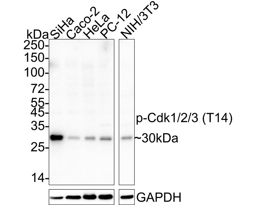
Western blot analysis of Phospho-Cdk1/2/3 (T14) on different lysates with Rabbit anti-Phospho-Cdk1/2/3 (T14) antibody at 1/1,000 dilution. Lane 1: SiHa cell lysate, Lane 2: Caco-2 cell lysate, Lane 3: HeLa cell lysate, Lane 4: PC-12 cell lysate, Lane 5: NIH/3T3 cell lysate, Lysates/proteins at 20 µg/Lane. Exposure time: 43 seconds; 4-20% SDS-PAGE gel. Proteins were transferred to a PVDF membrane and blocked with 5% NFDM/TBST for 1 hour at room temperature. The primary antibody at 1/1,000 dilution was used in 5% NFDM/TBST at 4℃ overnight. Goat Anti-Rabbit IgG - HRP Secondary Antibody at 1/50,000 dilution was used for 1 hour at room temperature.IHC
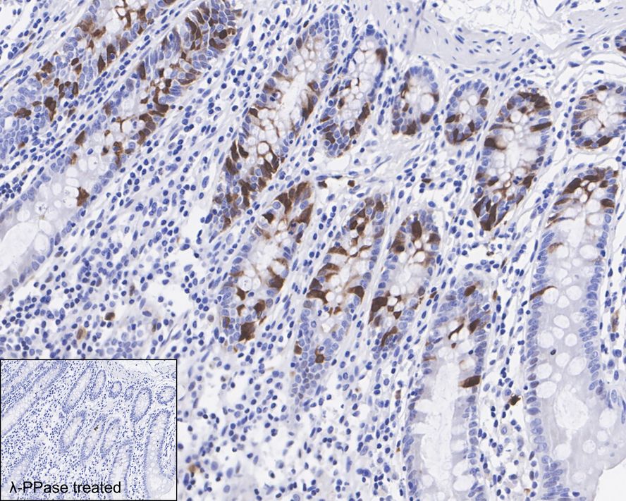
Immunohistochemical analysis of paraffin-embedded human colon tissue untreated / treated with λpp with Rabbit anti-Phospho-Cdk1/2/3 (T14) antibody at 1/1,000 dilution. The section was pre-treated using heat mediated antigen retrieval with sodium citrate buffer (pH 6.0) for 2 minutes. The tissues were blocked in 1% BSA for 20 minutes at room temperature, washed with ddH2O and PBS, and then probed with the primary antibody at 1/1,000 dilution for 1 hour at room temperature. The detection was performed using an HRP conjugated compact polymer system. DAB was used as the chromogen. Tissues were counterstained with hematoxylin and mounted with DPX.IHC
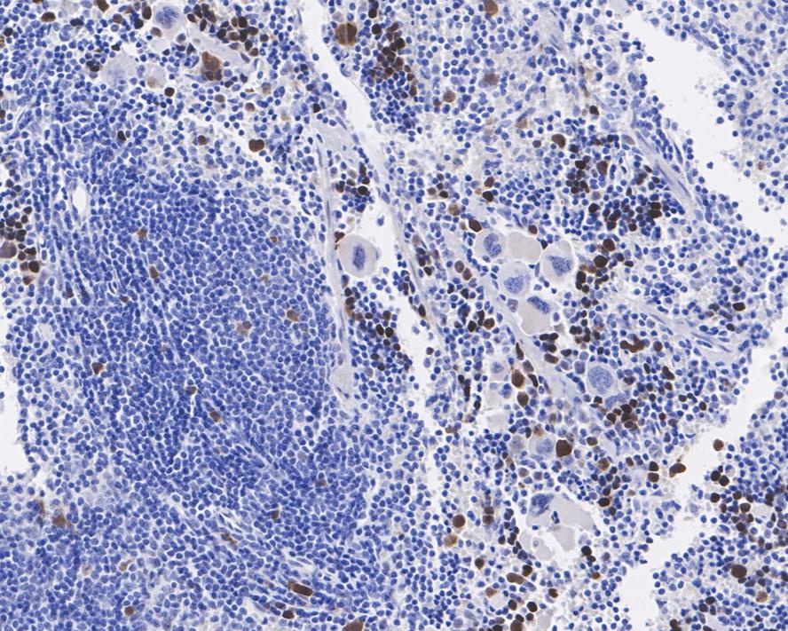
Immunohistochemical analysis of paraffin-embedded mouse spleen tissue with Rabbit anti-Phospho-Cdk1/2/3 (T14) antibody at 1/1,000 dilution. The section was pre-treated using heat mediated antigen retrieval with sodium citrate buffer (pH 6.0) for 2 minutes. The tissues were blocked in 1% BSA for 20 minutes at room temperature, washed with ddH2O and PBS, and then probed with the primary antibody at 1/1,000 dilution for 1 hour at room temperature. The detection was performed using an HRP conjugated compact polymer system. DAB was used as the chromogen. Tissues were counterstained with hematoxylin and mounted with DPX.IHC
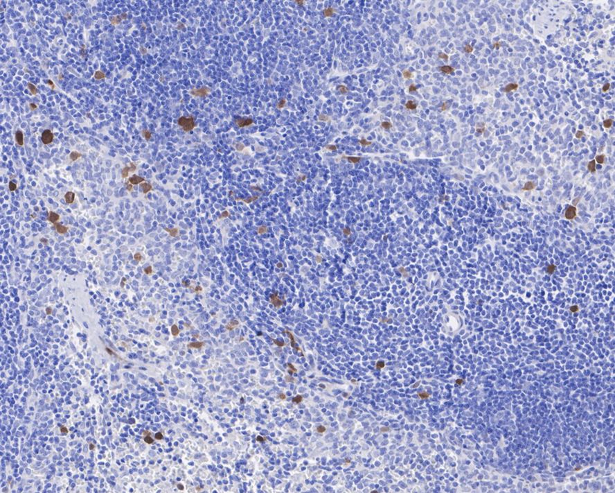
Immunohistochemical analysis of paraffin-embedded rat spleen tissue with Rabbit anti-Phospho-Cdk1/2/3 (T14) antibody at 1/1,000 dilution. The section was pre-treated using heat mediated antigen retrieval with sodium citrate buffer (pH 6.0) for 2 minutes. The tissues were blocked in 1% BSA for 20 minutes at room temperature, washed with ddH2O and PBS, and then probed with the primary antibody at 1/1,000 dilution for 1 hour at room temperature. The detection was performed using an HRP conjugated compact polymer system. DAB was used as the chromogen. Tissues were counterstained with hematoxylin and mounted with DPX.| Product Name | Phospho-Cdk1/2/3 (T14) Recombinant Rabbit Monoclonal Antibody |
|---|---|
| Antibody Type | Primary Antibodies |
| Immunogen | Synthetic phosphopeptide corresponding to residues surrounding Thr14 of human Cdk1 protein (P06493). |
| Modification | P-T14 |
| Clonality | monoclonal |
|---|---|
| Isotype | IgG |
| Host Species | Rabbit |
| Tested Applications | IHCWB |
| WB:1:1000 IHC:1:1000 |
|
| Species Reactivity | HumanMouseRat |
| Concentration | 1mg/ml |
| Purification | Protein A |
| Gene Symbol | CDK1,CDK2,CDK3 |
|---|---|
| Gene Synonyms | CDC2, CDC28A, P34CDC2 |
| Gene Full Name | cyclin dependent kinase 1,cyclin dependent kinase 2,cyclin dependent kinase 3 |
| Alternative Names | Cdc 2 antibody Cdc2 antibody CDC28A antibody CDK 1 antibody CDK1 antibody CDK1_HUMAN antibody CDKN1 antibody CELL CYCLE CONTROLLER CDC2 antibody Cell division control protein 2 antibody Cell division control protein 2 homolog antibody Cdc 2 antibody Cdc2 antibody CDC28A antibody CDK 1 antibody CDK1 antibody CDK1_HUMAN antibody CDKN1 antibody CELL CYCLE CONTROLLER CDC2 antibody Cell division control protein 2 antibody Cell division control protein 2 homolog antibody Cell division cycle 2 G1 to S and G2 to M antibody Cell division protein kinase 1 antibody Cell Divsion Cycle 2 Protein antibody Cyclin Dependent Kinase 1 antibody Cyclin-dependent kinase 1 antibody DKFZp686L20222 antibody MGC111195 antibody p34 Cdk1 antibody p34 protein kinase antibody P34CDC2 antibody Cdc2 related protein kinase antibody cdc2-related protein kinase antibody CDC28 antibody CDC2A antibody Cdk 2 antibody CDK2 antibody CDK2_HUMAN antibody CDKN2 antibody Cell devision kinase 2 antibody Cell division protein kinase 2 antibody Cyclin dependent kinase 2 antibody cyclin dependent kinase 2-alpha antibody Cyclin-dependent kinase 2 antibody kinase Cdc2 antibody MPF antibody p33 protein kinase antibody p33(CDK2) antibody Cdk 3 antibody Cdk3 antibody CDK3_HUMAN antibody CDKN3 antibody Cell division kinase 3 antibody Cell division protein kinase 3 antibody Cyclin dependent kinase 3 antibody Cyclin-dependent kinase 3 antibody OTTHUMP00000206828 antibody p36 antibody |
| Molecular Weight(MW) | 34kDa(Observed band size: 30kDa) |
| Function | Cyclin-dependent kinases (CDKs) are a predominant group of serine/threonine protein kinases involved in the regulation of the cell cycle and its progression, ensuring the integrity and functionality of cellular machinery. These regulatory enzymes play a crucial role in the regulation of eukaryotic cell cycle and transcription, as well as DNA repair, metabolism, and epigenetic regulation, in response to several extracellular and intracellular signals. They are present in all known eukaryotes, and their regulatory function in the cell cycle has been evolutionarily conserved. The catalytic activities of CDKs are regulated by interactions with CDK inhibitors (CKIs) and regulatory subunits known as cyclins. Cyclins have no enzymatic activity themselves, but they become active once they bind to CDKs. Without cyclin, CDK is less active than in the cyclin-CDK heterodimer complex. CDKs phosphorylate proteins on serine (S) or threonine (T) residues. The specificity of CDKs for their substrates is defined by the S/T-P-X-K/R sequence, where S/T is the phosphorylation site, P is proline, X is any amino acid, and the sequence ends with lysine (K) or arginine (R). This motif ensures CDKs accurately target and modify proteins, crucial for regulating cell cycle and other functions. Deregulation of the CDK activity is linked to various pathologies, including cancer, neurodegenerative diseases, and stroke. |
| Cellular Localization | Nucleus, Cytoplasm, Mitochondrion. |
WB
Western blot analysis of Phospho-Cdk1/2/3 (T14) on different lysates with Rabbit anti-Phospho-Cdk1/2/3 (T14) antibody at 1/1,000 dilution. Lane 1: SiHa cell lysate, Lane 2: Caco-2 cell lysate, Lane 3: HeLa cell lysate, Lane 4: PC-12 cell lysate, Lane 5: NIH/3T3 cell lysate, Lysates/proteins at 20 µg/Lane. Exposure time: 43 seconds; 4-20% SDS-PAGE gel. Proteins were transferred to a PVDF membrane and blocked with 5% NFDM/TBST for 1 hour at room temperature. The primary antibody at 1/1,000 dilution was used in 5% NFDM/TBST at 4℃ overnight. Goat Anti-Rabbit IgG - HRP Secondary Antibody at 1/50,000 dilution was used for 1 hour at room temperature.
IHC
Immunohistochemical analysis of paraffin-embedded human colon tissue untreated / treated with λpp with Rabbit anti-Phospho-Cdk1/2/3 (T14) antibody at 1/1,000 dilution. The section was pre-treated using heat mediated antigen retrieval with sodium citrate buffer (pH 6.0) for 2 minutes. The tissues were blocked in 1% BSA for 20 minutes at room temperature, washed with ddH2O and PBS, and then probed with the primary antibody at 1/1,000 dilution for 1 hour at room temperature. The detection was performed using an HRP conjugated compact polymer system. DAB was used as the chromogen. Tissues were counterstained with hematoxylin and mounted with DPX.
IHC
Immunohistochemical analysis of paraffin-embedded mouse spleen tissue with Rabbit anti-Phospho-Cdk1/2/3 (T14) antibody at 1/1,000 dilution. The section was pre-treated using heat mediated antigen retrieval with sodium citrate buffer (pH 6.0) for 2 minutes. The tissues were blocked in 1% BSA for 20 minutes at room temperature, washed with ddH2O and PBS, and then probed with the primary antibody at 1/1,000 dilution for 1 hour at room temperature. The detection was performed using an HRP conjugated compact polymer system. DAB was used as the chromogen. Tissues were counterstained with hematoxylin and mounted with DPX.
IHC
Immunohistochemical analysis of paraffin-embedded rat spleen tissue with Rabbit anti-Phospho-Cdk1/2/3 (T14) antibody at 1/1,000 dilution. The section was pre-treated using heat mediated antigen retrieval with sodium citrate buffer (pH 6.0) for 2 minutes. The tissues were blocked in 1% BSA for 20 minutes at room temperature, washed with ddH2O and PBS, and then probed with the primary antibody at 1/1,000 dilution for 1 hour at room temperature. The detection was performed using an HRP conjugated compact polymer system. DAB was used as the chromogen. Tissues were counterstained with hematoxylin and mounted with DPX.| Application Notes | WB:1:1000 IHC:1:1000 |
|---|
| Form | Liquid |
|---|---|
| Storage Instructions | Store at +4℃ after thawing. Aliquot store at -20℃. Avoid repeated freeze / thaw cycles. |
| Storage Buffer | 1*TBS (pH7.4), 0.05% BSA, 40% Glycerol. Preservative: 0.05% Sodium Azide. |


