IHC
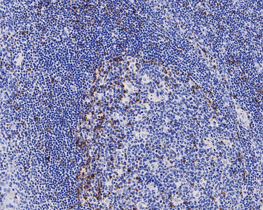
Immunohistochemical analysis of paraffin-embedded human tonsils tissue with Rabbit anti-CTLA-4 antibody at 1/1,000 dilution. The section was pre-treated using heat mediated antigen retrieval with Tris-EDTA buffer (pH 9.0) for 20 minutes. The tissues were blocked in 1% BSA for 20 minutes at room temperature, washed with ddH2O and PBS, and then probed with the primary antibody at 1/1,000 dilution for 1 hour at room temperature. The detection was performed using an HRP conjugated compact polymer system. DAB was used as the chromogen. Tissues were counterstained with hematoxylin and mounted with DPX.IHC
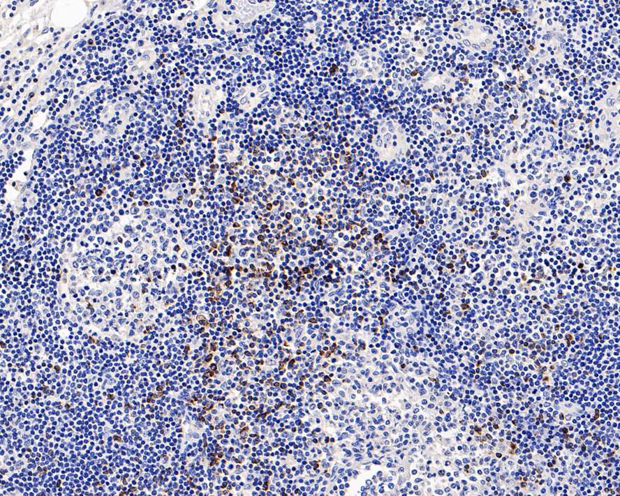
Immunohistochemical analysis of paraffin-embedded human lymph nodes tissue with Rabbit anti-CTLA-4 antibody at 1/1,000 dilution. The section was pre-treated using heat mediated antigen retrieval with Tris-EDTA buffer (pH 9.0) for 20 minutes. The tissues were blocked in 1% BSA for 20 minutes at room temperature, washed with ddH2O and PBS, and then probed with the primary antibody at 1/1,000 dilution for 1 hour at room temperature. The detection was performed using an HRP conjugated compact polymer system. DAB was used as the chromogen. Tissues were counterstained with hematoxylin and mounted with DPX.mIHC
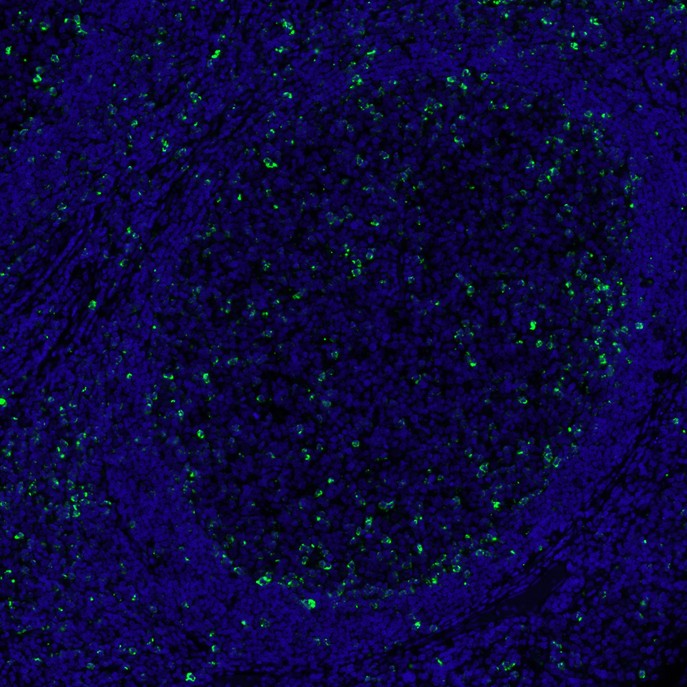
mIHC analysis of human tonsils tissue (Formalin/PFA-fixed paraffin-embedded sections) with Rabbit anti-CTLA-4 antibody at 1/100 dilution. The immunostaining was performed with the HyperView mTSA Kit. Heat mediated antigen retrieval with Tris-EDTA buffer (pH 9.0) for 30 mins at 95℃. DAPI (blue) was used as a nuclear counter stain. Image acquisition was performed with Olympus VS200 Slide Scanner.| Product Name | CTLA-4 Recombinant Rabbit Monoclonal Antibody |
|---|---|
| Antibody Type | Primary Antibodies |
| Immunogen | Synthetic peptide within Human CTLA4 aa 150 to the C-terminus. |
| Clonality | monoclonal |
|---|---|
| Isotype | IgG |
| Host Species | Rabbit |
| Tested Applications | IHC |
| IHC:1:200-1:1000 mIHC:1:500 |
|
| Species Reactivity | HumanMouse |
| Concentration | 1mg/ml |
| Purification | Protein A |
| Gene Symbol | CTLA4 |
|---|---|
| Gene Synonyms | CD GSE GRD4 ALPS5 CD152 CTLA-4 IDDM12 CELIAC3 |
| Gene Full Name | cytotoxic T-lymphocyte associated protein 4 |
| Gene Summary | This gene is a member of the immunoglobulin superfamily and encodes a protein which transmits an inhibitory signal to T cells. The protein contains a V domain, a transmembrane domain, and a cytoplasmic tail. Alternate transcriptional splice variants, encoding different isoforms, have been characterized. The membrane-bound isoform functions as a homodimer interconnected by a disulfide bond, while the soluble isoform functions as a monomer. Mutations in this gene have been associated with insulin-dependent diabetes mellitus, Graves disease, Hashimoto thyroiditis, celiac disease, systemic lupus erythematosus, thyroid-associated orbitopathy, and other autoimmune diseases. [provided by RefSeq, Jul 2008] |
| Alternative Names | ALPS5 antibody CD 152 antibody CD antibody CD152 antibody CD152 antigen antibody CD152 isoform antibody Celiac disease 3 antibody CELIAC3 antibody CTLA 4 antibody CTLA-4 antibody ALPS5 antibody CD 152 antibody CD antibody CD152 antibody CD152 antigen antibody CD152 isoform antibody Celiac disease 3 antibody CELIAC3 antibody CTLA 4 antibody CTLA-4 antibody CTLA4 antibody CTLA4_HUMAN antibody Cytotoxic T cell associated 4 antibody Cytotoxic T lymphocyte antigen 4 antibody Cytotoxic T lymphocyte associated 4 antibody Cytotoxic T lymphocyte associated 4 soluble isoform included antibody Cytotoxic T lymphocyte associated antigen 4 antibody Cytotoxic T lymphocyte associated antigen 4 short spliced form antibody Cytotoxic T lymphocyte associated protein 4 antibody Cytotoxic T lymphocyte associated serine esterase 4 antibody Cytotoxic T lymphocyte protein 4 antibody Cytotoxic T-lymphocyte protein 4 antibody Cytotoxic T-lymphocyte-associated antigen 4 antibody GRD4 antibody GSE antibody ICOS antibody IDDM12 antibody insulin-dependent diabetes mellitus 12 antibody Ligand and transmembrane spliced cytotoxic T lymphocyte associated antigen 4 antibody OTTHUMP00000216623 antibody |
| Molecular Weight(MW) | 25kDa |
| Cellular Localization | Cell membrane. |
IHC
Immunohistochemical analysis of paraffin-embedded human tonsils tissue with Rabbit anti-CTLA-4 antibody at 1/1,000 dilution. The section was pre-treated using heat mediated antigen retrieval with Tris-EDTA buffer (pH 9.0) for 20 minutes. The tissues were blocked in 1% BSA for 20 minutes at room temperature, washed with ddH2O and PBS, and then probed with the primary antibody at 1/1,000 dilution for 1 hour at room temperature. The detection was performed using an HRP conjugated compact polymer system. DAB was used as the chromogen. Tissues were counterstained with hematoxylin and mounted with DPX.
IHC
Immunohistochemical analysis of paraffin-embedded human lymph nodes tissue with Rabbit anti-CTLA-4 antibody at 1/1,000 dilution. The section was pre-treated using heat mediated antigen retrieval with Tris-EDTA buffer (pH 9.0) for 20 minutes. The tissues were blocked in 1% BSA for 20 minutes at room temperature, washed with ddH2O and PBS, and then probed with the primary antibody at 1/1,000 dilution for 1 hour at room temperature. The detection was performed using an HRP conjugated compact polymer system. DAB was used as the chromogen. Tissues were counterstained with hematoxylin and mounted with DPX.
mIHC
mIHC analysis of human tonsils tissue (Formalin/PFA-fixed paraffin-embedded sections) with Rabbit anti-CTLA-4 antibody at 1/100 dilution. The immunostaining was performed with the HyperView mTSA Kit. Heat mediated antigen retrieval with Tris-EDTA buffer (pH 9.0) for 30 mins at 95℃. DAPI (blue) was used as a nuclear counter stain. Image acquisition was performed with Olympus VS200 Slide Scanner.| Application Notes | IHC:1:200-1:1000 mIHC:1:500 |
|---|
| Form | Liquid |
|---|---|
| Storage Instructions | Store at +4℃ after thawing. Aliquot store at -20℃. Avoid repeated freeze / thaw cycles. |
| Storage Buffer | PBS (pH7.4), 0.1% BSA, 40% Glycerol. Preservative: 0.05% Sodium Azide. |


