WB
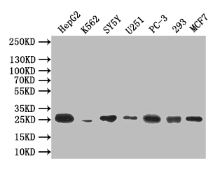
Western Blot: Positive WB detected in: HepG2 whole cell lysate, K562 whole cell lysate, SY5Y whole cell lysate, U251 whole cell lysate, PC-3 whole cell lysate, 293 whole cell lysate, MCF7 whole cell lysate. All lanes: CTLA4 antibody at 1:500. Secondary: Goat polyclonal to mouse IgG at 1/50000 dilution.ICC/IF
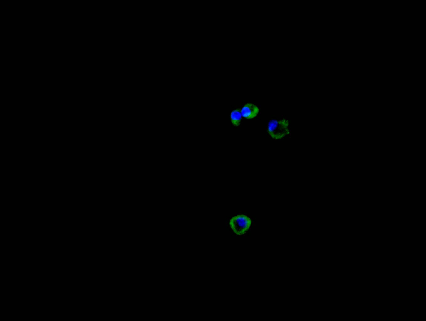
Immunofluorescence staining of HEPG2 cell with CTLA4 Recombinant Monoclonal Antibody at 1:30, counter-stained with DAPI. The cells were fixed in 4% formaldehyde and blocked in 10% normal Goat Serum. The cells were then incubated with the antibody overnight at 4C. The secondary antibody was FITC-conjugated AffiniPure Goat Anti-Mouse IgG(H+L).FC
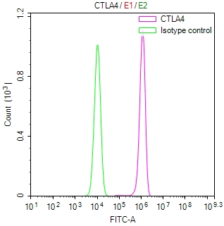
Overlay Peak curve showing JK cells surface stained with CTLA4 Recombinant Monoclonal Antibody (red line) at 1:50. Then 10% normal goat serum to block non-specific protein-protein interactions followed by the antibody (1ug/1*106cells) for 45min at 4℃. The secondary antibody used was FITC-conjugated Goat Anti-Mouse IgG(H+L) at 1/200 dilution for 35 min at 4°C. Isotype control antibody (green line) was mouse IgG1 (1µg/1*106cells) used under the same conditions. Acquisition of >10,015 events was performed.| Product Name | CTLA4 Recombinant Monoclonal Antibody |
|---|---|
| Antibody Type | Primary Antibodies |
| Immunogen | Recombinant Human CTLA4 protein |
| Clonality | Monoclonal |
|---|---|
| Isotype | IgG2a |
| Host Species | Mouse |
| Tested Applications | FCICC/IFWB |
| WB:1:200-1:2000 ICC/IF:1:50-1:200 |
|
| Species Reactivity | Human |
| Concentration | 1mg/ml |
| Purification | Affinity purified |
| Gene Symbol | CTLA4 |
|---|---|
| Gene Synonyms | CD GSE GRD4 ALPS5 CD152 CTLA-4 IDDM12 CELIAC3 |
| Gene Full Name | cytotoxic T-lymphocyte associated protein 4 |
| Gene Summary | This gene is a member of the immunoglobulin superfamily and encodes a protein which transmits an inhibitory signal to T cells. The protein contains a V domain, a transmembrane domain, and a cytoplasmic tail. Alternate transcriptional splice variants, encoding different isoforms, have been characterized. The membrane-bound isoform functions as a homodimer interconnected by a disulfide bond, while the soluble isoform functions as a monomer. Mutations in this gene have been associated with insulin-dependent diabetes mellitus, Graves disease, Hashimoto thyroiditis, celiac disease, systemic lupus erythematosus, thyroid-associated orbitopathy, and other autoimmune diseases. [provided by RefSeq, Jul 2008] |
| Alternative Names | ALPS5 antibody CD 152 antibody CD antibody CD152 antibody CD152 antigen antibody CD152 isoform antibody Celiac disease 3 antibody CELIAC3 antibody CTLA 4 antibody CTLA-4 antibody CTLA4 antibody CTLA4_HUMAN antibody Cytotoxic T cell associated 4 antibody Cytotoxic T lymphocyte antigen 4 antibody Cytotoxic T lymphocyte associated 4 antibody Cytotoxic T lymphocyte associated 4 soluble isoform included antibody Cytotoxic T lymphocyte associated antigen 4 antibody Cytotoxic T lymphocyte associated antigen 4 short spliced form antibody Cytotoxic T lymphocyte associated protein 4 antibody Cytotoxic T lymphocyte associated serine esterase 4 antibody Cytotoxic T lymphocyte protein 4 antibody Cytotoxic T-lymphocyte protein 4 antibody Cytotoxic T-lymphocyte-associated antigen 4 antibody GRD4 antibody GSE antibody ICOS antibody IDDM12 antibody insulin-dependent diabetes mellitus 12 antibody Ligand and transmembrane spliced cytotoxic T lymphocyte associated antigen 4 antibody OTTHUMP00000216623 antibody |
| Molecular Weight(MW) | 25 kDa |
| Cellular Localization | Cell membrane; Single-pass type I membrane protein. Note=Exists primarily an intracellular antigen whose surface expression is tightly regulated by restricted trafficking to the cell surface and rapid internalization. |
WB
Western Blot: Positive WB detected in: HepG2 whole cell lysate, K562 whole cell lysate, SY5Y whole cell lysate, U251 whole cell lysate, PC-3 whole cell lysate, 293 whole cell lysate, MCF7 whole cell lysate. All lanes: CTLA4 antibody at 1:500. Secondary: Goat polyclonal to mouse IgG at 1/50000 dilution.
ICC/IF
Immunofluorescence staining of HEPG2 cell with CTLA4 Recombinant Monoclonal Antibody at 1:30, counter-stained with DAPI. The cells were fixed in 4% formaldehyde and blocked in 10% normal Goat Serum. The cells were then incubated with the antibody overnight at 4C. The secondary antibody was FITC-conjugated AffiniPure Goat Anti-Mouse IgG(H+L).
FC
Overlay Peak curve showing JK cells surface stained with CTLA4 Recombinant Monoclonal Antibody (red line) at 1:50. Then 10% normal goat serum to block non-specific protein-protein interactions followed by the antibody (1ug/1*106cells) for 45min at 4℃. The secondary antibody used was FITC-conjugated Goat Anti-Mouse IgG(H+L) at 1/200 dilution for 35 min at 4°C. Isotype control antibody (green line) was mouse IgG1 (1µg/1*106cells) used under the same conditions. Acquisition of >10,015 events was performed.| Application Notes | WB:1:200-1:2000 ICC/IF:1:50-1:200 |
|---|
| Form | Liquid |
|---|---|
| Storage Instructions | Upon receipt, store at -20°C or -80°C. Avoid repeated freeze. |
| Storage Buffer | 0.03% Proclin 300 Constituents: 50% Glycerol, 0.01M PBS, PH 7.4 |


