WB
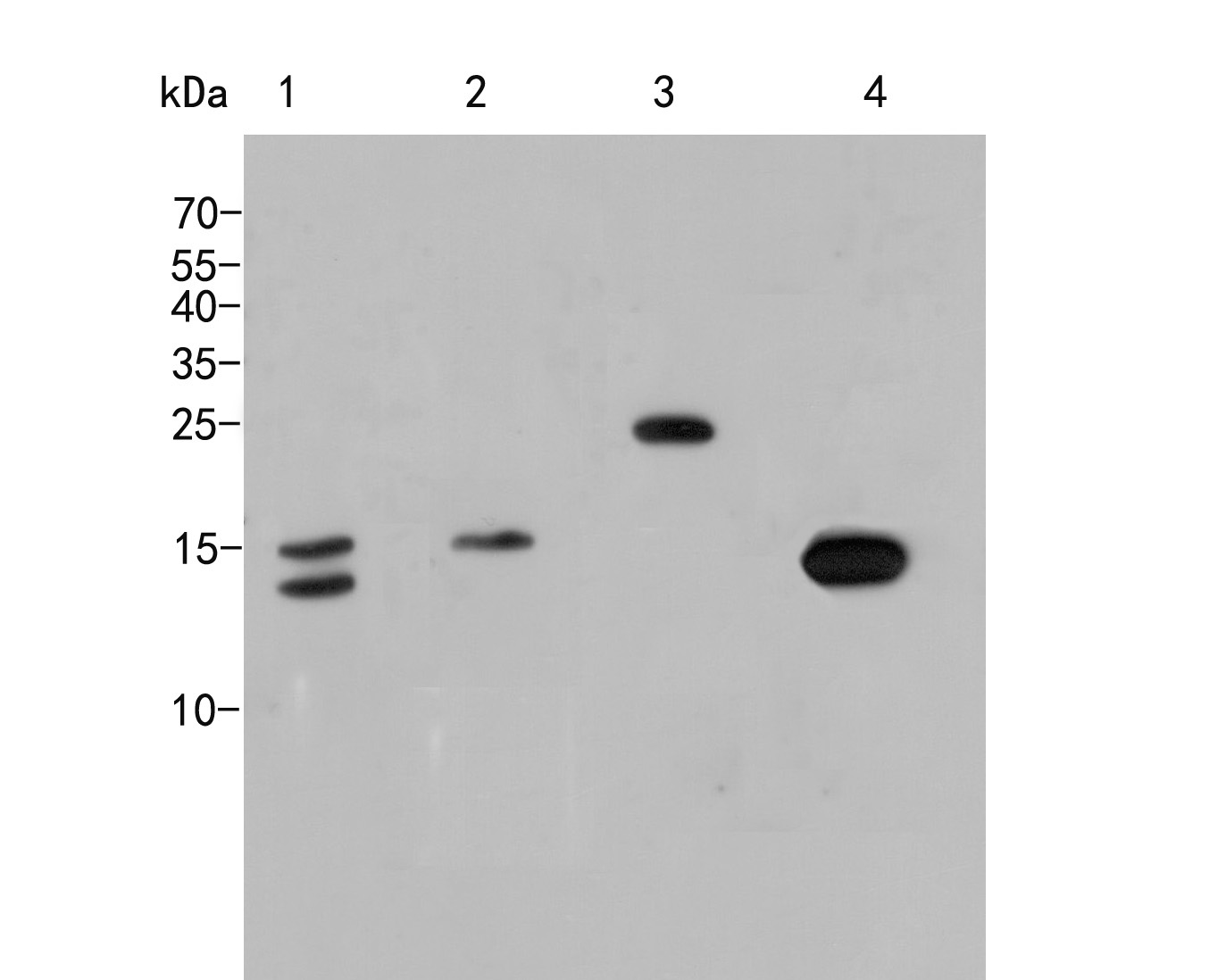
Western blot analysis of Histone H3 on different lysates. Proteins were transferred to a PVDF membrane and blocked with 5% NFDM/TBST for 1 hour at room temperature. The primary antibody (1/1000) was used in 5% NFDM/TBST at room temperature for 2 hours. Goat Anti-Rabbit IgG - HRP Secondary Antibody at 1:5,000 dilution was used for 1 hour at room temperature. Positive control: Lane 1: Hela cell lysate, Lane 2: NIH/3T3 cell lysate, Lane 3: Rice tissue lysate, Lane 4: Zebrafish tissue cell lysate.IHC
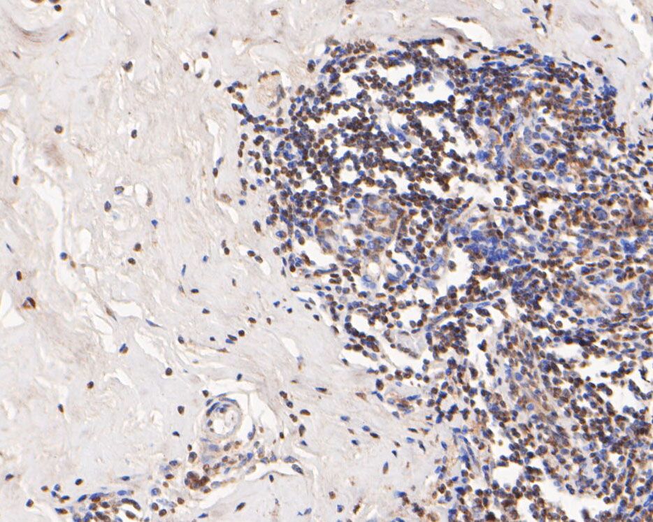
Immunohistochemical analysis of paraffin-embedded human breast cancer tissue using anti-Histone H3 antibody. The section was pre-treated using heat mediated antigen retrieval with sodium citrate buffer (pH 6.0) (high pressure) for 2 minutes. The tissues were blocked in 5% BSA for 30 minutes at room temperature, washed with ddH2O and PBS, and then probed with the primary antibody (1/400) for 30 minutes at room temperature. The detection was performed using an HRP conjugated compact polymer system. DAB was used as the chromogen. Tissues were counterstained with hematoxylin and mounted with DPX.IHC
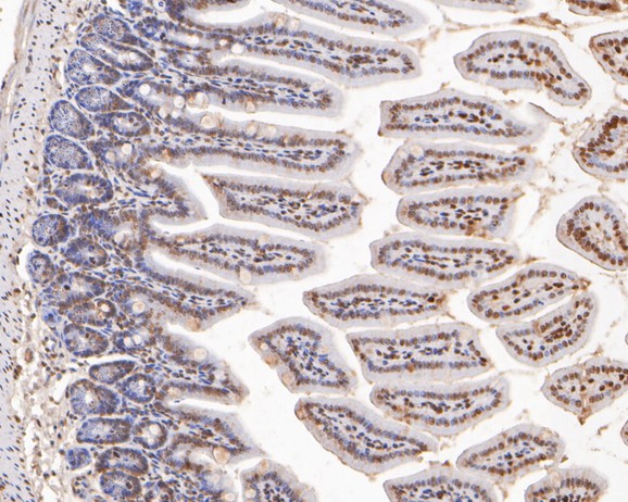
Immunohistochemical analysis of paraffin-embedded mouse colon cancer tissue using anti-Histone H3 antibody. The section was pre-treated using heat mediated antigen retrieval with sodium citrate buffer (pH 6.0) (high pressure) for 2 minutes. The tissues were blocked in 5% BSA for 30 minutes at room temperature, washed with ddH2O and PBS, and then probed with the primary antibody (1/400) for 30 minutes at room temperature. The detection was performed using an HRP conjugated compact polymer system. DAB was used as the chromogen. Tissues were counterstained with hematoxylin and mounted with DPX.| Product Name | Histone H3 Rabbit Polyclonal Antibody |
|---|---|
| Antibody Type | Primary Antibodies |
| Immunogen | Synthetic peptide within Zebrafish Histone H3 aa 1-50 / 136. |
| Clonality | Polyclonal |
|---|---|
| Isotype | IgG |
| Host Species | Rabbit |
| Tested Applications | IHCWB |
| WB:1:500-1:2000 IHC:1:400 |
|
| Species Reactivity | HumanMouseZebrafish |
| Concentration | 1mg/ml |
| Purification | Protein A |
| Alternative Names | H3 histone family member E pseudogene antibody H3 histone family member A antibody H3/A antibody H31_HUMAN antibody H3F3 antibody H3FA antibody Hist1h3a antibody HIST1H3B antibody HIST1H3C antibody HIST1H3D antibody H3 histone family member E pseudogene antibody H3 histone family member A antibody H3/A antibody H31_HUMAN antibody H3F3 antibody H3FA antibody Hist1h3a antibody HIST1H3B antibody HIST1H3C antibody HIST1H3D antibody HIST1H3E antibody HIST1H3F antibody HIST1H3G antibody HIST1H3H antibody HIST1H3I antibody HIST1H3J antibody HIST3H3 antibody histone 1 H3a antibody Histone cluster 1 H3a antibody Histone H3 3 pseudogene antibody Histone H3.1 antibody Histone H3/a antibody Histone H3/b antibody Histone H3/c antibody Histone H3/d antibody Histone H3/f antibody Histone H3/h antibody Histone H3/i antibody Histone H3/j antibody Histone H3/k antibody Histone H3/l antibody |
|---|---|
| Molecular Weight(MW) | 15kDa |
| Function | Variant histone H3 which replaces conventional H3 in a wide range of nucleosomes in active genes. Constitutes the predominant form of histone H3 in non-dividing cells and is incorporated into chromatin independently of DNA synthesis. Deposited at sites of nucleosomal displacement throughout transcribed genes, suggesting that it represents an epigenetic imprint of transcriptionally active chromatin. Nucleosomes wrap and compact DNA into chromatin, limiting DNA accessibility to the cellular machineries which require DNA as a template. Histones thereby play a central role in transcription regulation, DNA repair, DNA replication and chromosomal stability. DNA accessibility is regulated via a complex set of post-translational modifications of histones, also called histone code, and nucleosome remodeling. |
| Cellular Localization | Nucleus, Chromosome. |
WB
Western blot analysis of Histone H3 on different lysates. Proteins were transferred to a PVDF membrane and blocked with 5% NFDM/TBST for 1 hour at room temperature. The primary antibody (1/1000) was used in 5% NFDM/TBST at room temperature for 2 hours. Goat Anti-Rabbit IgG - HRP Secondary Antibody at 1:5,000 dilution was used for 1 hour at room temperature. Positive control: Lane 1: Hela cell lysate, Lane 2: NIH/3T3 cell lysate, Lane 3: Rice tissue lysate, Lane 4: Zebrafish tissue cell lysate.
IHC
Immunohistochemical analysis of paraffin-embedded human breast cancer tissue using anti-Histone H3 antibody. The section was pre-treated using heat mediated antigen retrieval with sodium citrate buffer (pH 6.0) (high pressure) for 2 minutes. The tissues were blocked in 5% BSA for 30 minutes at room temperature, washed with ddH2O and PBS, and then probed with the primary antibody (1/400) for 30 minutes at room temperature. The detection was performed using an HRP conjugated compact polymer system. DAB was used as the chromogen. Tissues were counterstained with hematoxylin and mounted with DPX.
IHC
Immunohistochemical analysis of paraffin-embedded mouse colon cancer tissue using anti-Histone H3 antibody. The section was pre-treated using heat mediated antigen retrieval with sodium citrate buffer (pH 6.0) (high pressure) for 2 minutes. The tissues were blocked in 5% BSA for 30 minutes at room temperature, washed with ddH2O and PBS, and then probed with the primary antibody (1/400) for 30 minutes at room temperature. The detection was performed using an HRP conjugated compact polymer system. DAB was used as the chromogen. Tissues were counterstained with hematoxylin and mounted with DPX.| Application Notes | WB:1:500-1:2000 IHC:1:400 |
|---|
| Form | Liquid |
|---|---|
| Storage Instructions | Store at +4℃ after thawing. Aliquot store at -20℃. Avoid repeated freeze / thaw cycles. |
| Storage Buffer | PBS (pH7.4), 0.1% BSA, 40% Glycerol. Preservative: 0.05% Sodium Azide. |


