WB
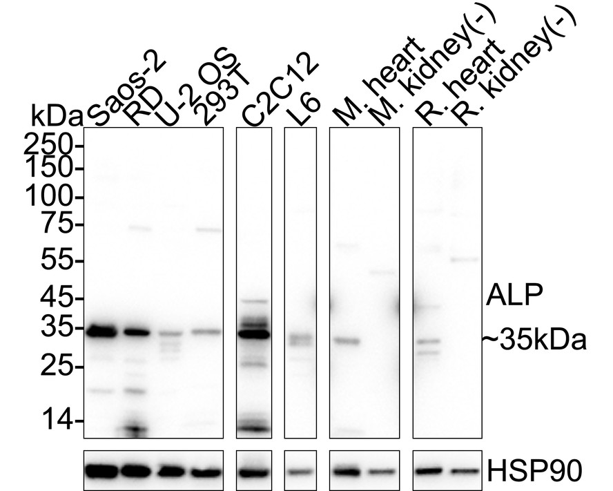
Western blot analysis of ALP on different lysates with Rabbit anti-ALP antibody at 1/5,000 dilution. Lane 1: Saos-2 cell lysate, Lane 2: RD cell lysate, Lane 3: U-2 OS cell lysate, Lane 4: 293T cell lysate, Lane 5: C2C12 cell lysate, Lane 6: L6 cell lysate, Lane 7: Mouse heart tissue lysate, Lane 8: Mouse kidney tissue lysate (negative), Lane 9: Rat heart tissue lysate, Lane 10: Rat kidney tissue lysate (negative), Lysates/proteins at 20 µg/Lane. Exposure time: 10 seconds; 4-20% SDS-PAGE gel. Proteins were transferred to a PVDF membrane and blocked with 5% NFDM/TBST for 1 hour at room temperature. The primary antibody at 1/5,000 dilution was used in primary antibody dilution at 4℃ overnight. Goat Anti-Rabbit IgG - HRP Secondary Antibody at 1/50,000 dilution was used for 1 hour at room temperature.IP
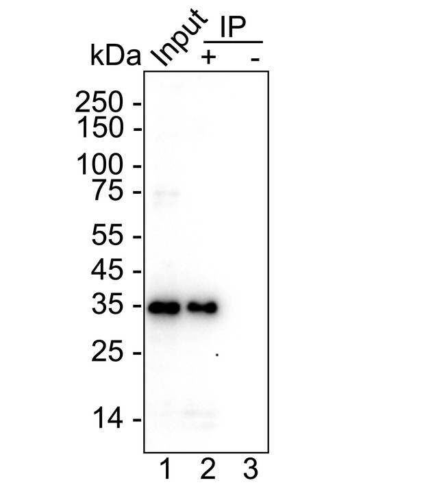
ALP was immunoprecipitated from 0.2 mg Saos-2 cell lysate with Rabbit anti-ALP antibody at 2 µg/10 µl beads. Western blot was performed from the immunoprecipitate using Rabbit anti-ALP antibody at 1/5,000 dilution. HRP Conjugated Anti-Rabbit IgG for IP Nano-secondary antibody at 1/5,000 dilution was used for 1 hour at room temperature. Lane 1: Saos-2 cell lysate (input), Lane 2: Rabbit anti-ALP antibody IP in Saos-2 cell lysate, Lane 3: Rabbit IgG instead of Rabbit anti-ALP antibody in Saos-2 cell lysate, Blocking/Dilution buffer: 5% NFDM/TBST, Exposure time: 46 seconds.| Product Name | ALP Recombinant Rabbit Monoclonal Antibody |
|---|---|
| Antibody Type | Primary Antibodies |
| Immunogen | Recombinant protein within human ALP aa 1-364. |
| Clonality | monoclonal |
|---|---|
| Isotype | IgG |
| Host Species | Rabbit |
| Tested Applications | IPWB |
| WB:1:5000 IP:1-2μg/sample |
|
| Species Reactivity | HumanMouseRat |
| Concentration | 1mg/ml |
| Purification | Protein A |
| Gene Symbol | PDLIM3 |
|---|---|
| Gene Synonyms | ALP |
| Gene Full Name | PDZ and LIM domain 3 |
| Gene Summary | The protein encoded by this gene contains a PDZ domain and a LIM domain, indicating that it may be involved in cytoskeletal assembly. In support of this, the encoded protein has been shown to bind the spectrin-like repeats of alpha-actinin-2 and to colocalize with alpha-actinin-2 at the Z lines of skeletal muscle. Alternatively spliced transcript variants encoding multiple isoforms have been observed for this gene. Aberrant alternative splicing of this gene may play a role in myotonic dystrophy. [provided by RefSeq, Apr 2012] |
| Alternative Names | Actinin associated LIM protein antibody Actinin-associated LIM protein antibody Alpha actinin 2 associated LIM protein antibody Alpha-actinin-2-associated LIM protein antibody DKFZp686L0362 antibody Enigma homolog antibody PDLI3_HUMAN antibody PDLIM 3 antibody PDLIM3 antibody PDZ and LIM domain 3 antibody Actinin associated LIM protein antibody Actinin-associated LIM protein antibody Alpha actinin 2 associated LIM protein antibody Alpha-actinin-2-associated LIM protein antibody DKFZp686L0362 antibody Enigma homolog antibody PDLI3_HUMAN antibody PDLIM 3 antibody PDLIM3 antibody PDZ and LIM domain 3 antibody PDZ and LIM domain protein 3 antibody |
| Molecular Weight(MW) | 39kDa(Observed band size: 35kDa) |
| Cellular Localization | Cytoplasm, myofibril, sarcomere, Z line. |
WB
Western blot analysis of ALP on different lysates with Rabbit anti-ALP antibody at 1/5,000 dilution. Lane 1: Saos-2 cell lysate, Lane 2: RD cell lysate, Lane 3: U-2 OS cell lysate, Lane 4: 293T cell lysate, Lane 5: C2C12 cell lysate, Lane 6: L6 cell lysate, Lane 7: Mouse heart tissue lysate, Lane 8: Mouse kidney tissue lysate (negative), Lane 9: Rat heart tissue lysate, Lane 10: Rat kidney tissue lysate (negative), Lysates/proteins at 20 µg/Lane. Exposure time: 10 seconds; 4-20% SDS-PAGE gel. Proteins were transferred to a PVDF membrane and blocked with 5% NFDM/TBST for 1 hour at room temperature. The primary antibody at 1/5,000 dilution was used in primary antibody dilution at 4℃ overnight. Goat Anti-Rabbit IgG - HRP Secondary Antibody at 1/50,000 dilution was used for 1 hour at room temperature.
IP
ALP was immunoprecipitated from 0.2 mg Saos-2 cell lysate with Rabbit anti-ALP antibody at 2 µg/10 µl beads. Western blot was performed from the immunoprecipitate using Rabbit anti-ALP antibody at 1/5,000 dilution. HRP Conjugated Anti-Rabbit IgG for IP Nano-secondary antibody at 1/5,000 dilution was used for 1 hour at room temperature. Lane 1: Saos-2 cell lysate (input), Lane 2: Rabbit anti-ALP antibody IP in Saos-2 cell lysate, Lane 3: Rabbit IgG instead of Rabbit anti-ALP antibody in Saos-2 cell lysate, Blocking/Dilution buffer: 5% NFDM/TBST, Exposure time: 46 seconds.| Application Notes | WB:1:5000 IP:1-2μg/sample |
|---|
| Form | Liquid |
|---|---|
| Storage Instructions | Store at +4℃ after thawing. Aliquot store at -20℃. Avoid repeated freeze / thaw cycles. |
| Storage Buffer | PBS (pH7.4), 0.1% BSA, 40% Glycerol. Preservative: 0.05% Sodium Azide. |


