WB
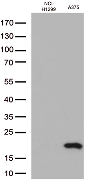
Western blot analysis of extracts (35ug) from 2 cell lines lysates by using anti-CTAG1B monoclonal antibody (1:500).IHC
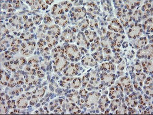
Immunohistochemical staining of paraffin-embedded Human pancreas tissue within the normal limits using anti-CTAG1B mouse monoclonal antibody. Heat-induced epitope retrieval by EDTA solution buffer pH 8.0 at 120°C for 3 min.IHC
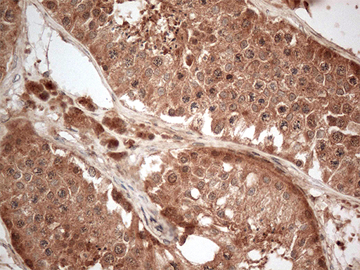
Immunohistochemical staining of paraffin-embedded Human testicle tissue within the normal limits using anti-CTAG1B mouse monoclonal antibody. Heat-induced epitope retrieval by EDTA solution buffer pH 8.0 at 120°C for 3 min.ICC/IF
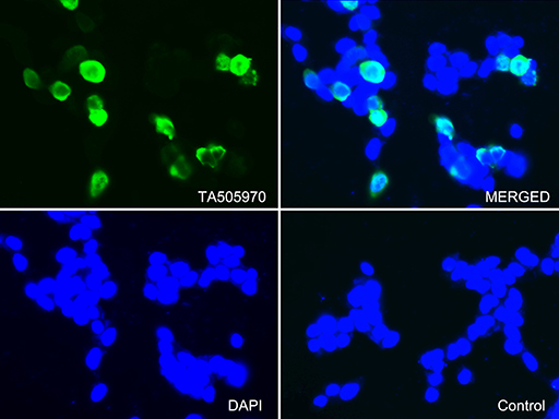
Immunofluorescent staining of 293T cells transfected by pCMV6-ENTRY CTAG1B using anti-CTAG1B antibody (green, upper left; DAPI/blue, lower left; MERGED, upper right). 293T cells transfected with empty vector served as a negative control (MERGED, lower right) (1:100).FC
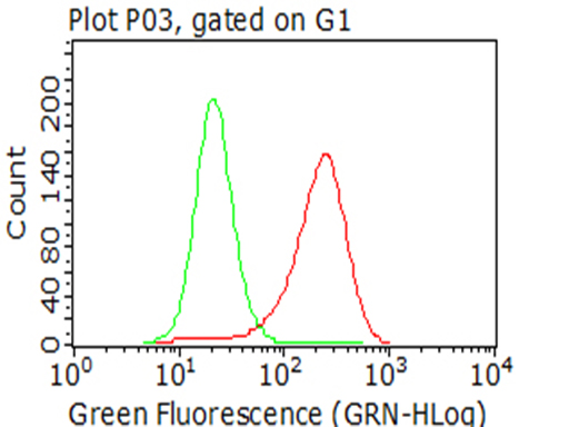
Flow cytometric analysis of living A375 cells, using anti-CTAG1B antibody (Red), compared to an isotype control (green) (1:100).| Product Name | NY-ESO-1 (CTAG1B) Mouse Monoclonal Antibody |
|---|---|
| Antibody Type | Primary Antibodies |
| Immunogen | Full length human recombinant protein of human CTAG1B(NP_001318) produced in HEK293T cell. |
| Clonality | monoclonal |
|---|---|
| Isotype | IgG1 |
| Host Species | Mouse |
| Tested Applications | FCICC/IFIHCWB |
| WB:1:2000 IHC:1:150 ICC/IF:1:100 FC:1:100 |
|
| Species Reactivity | Human |
| Concentration | 1mg/ml |
| Purification | Protein A/G |
| Gene Symbol | CTAG1B |
|---|---|
| Gene Synonyms | CTAG ESO1 CT6.1 CTAG1 LAGE-2 LAGE2B NY-ESO-1 |
| Gene Full Name | cancer/testis antigen 1B |
| Gene Summary | The protein encoded by this gene is an antigen that is overexpressed in many cancers but that is also expressed in normal testis. This gene is found in a duplicated region of the X-chromosome and therefore has a neighboring gene of identical sequence. [provided by RefSeq, Jan 2012] |
| Alternative Names | CT6.1 CTAG CTAG1 ESO1 LAGE-2 LAGE2B NY-ESO-1 |
| Molecular Weight(MW) | 18kDa |
| Cellular Localization | Cytoplasm. |
WB
Western blot analysis of extracts (35ug) from 2 cell lines lysates by using anti-CTAG1B monoclonal antibody (1:500).
IHC
Immunohistochemical staining of paraffin-embedded Human pancreas tissue within the normal limits using anti-CTAG1B mouse monoclonal antibody. Heat-induced epitope retrieval by EDTA solution buffer pH 8.0 at 120°C for 3 min.
IHC
Immunohistochemical staining of paraffin-embedded Human testicle tissue within the normal limits using anti-CTAG1B mouse monoclonal antibody. Heat-induced epitope retrieval by EDTA solution buffer pH 8.0 at 120°C for 3 min.
ICC/IF
Immunofluorescent staining of 293T cells transfected by pCMV6-ENTRY CTAG1B using anti-CTAG1B antibody (green, upper left; DAPI/blue, lower left; MERGED, upper right). 293T cells transfected with empty vector served as a negative control (MERGED, lower right) (1:100).
FC
Flow cytometric analysis of living A375 cells, using anti-CTAG1B antibody (Red), compared to an isotype control (green) (1:100).| Application Notes | WB:1:2000 IHC:1:150 ICC/IF:1:100 FC:1:100 |
|---|
| Form | Liquid |
|---|---|
| Storage Instructions | Store at -20°C as received. |
| Storage Buffer | PBS (pH 7.3) containing 1% BSA, 50% glycerol and 0.02% sodium azide. |


