WB
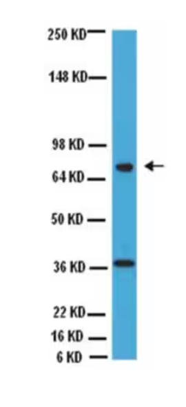
Western Blot Analysis: Representative lot data. NIH/3T3 lysate was resolved by electrophoresis, transferred to PVDF membrane and probed with anti-CHAT (1:1000 dilution). Proteins were visualized using a rabbit anti-goat secondary antibody conjugated to HRP and a chemiluminescence detection system.WB
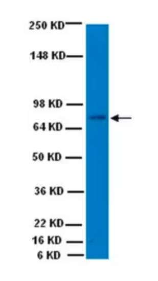
Western Blot Analysis: Mouse brain lysate was resolved by electrophoresis, transferred to PVDF membrane and probed with anti-CHAT (1:1000 dilution of this lot). Proteins were visualized using a rabbit anti-goat secondary antibody conjugated to HRP and a chemiluminescence detection system.IF-P
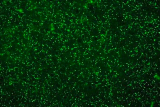
Coronal section of rat neocortex stained with CHAT antibody showing the choline acetyltransferase positive cholinergic nerve fibers and terminals.| Product Name | Goat polyclonal to Choline Acetyltransferase |
|---|---|
| Antibody Type | Primary Antibodies |
| Immunogen | Synthetic peptide |
| Clonality | Polyclonal |
|---|---|
| Isotype | IgG |
| Host Species | Goat |
| Tested Applications | IF-PWB |
| WB:1:1000 IF-P:1:100 |
|
| Species Reactivity | HumanMouseRat |
| Concentration | 0.5mg/ml |
| Purification | Affinity purified |
| Gene Symbol | CHAT |
|---|---|
| Gene Synonyms | CMS6 CMS1A CMS1A2 CHOACTASE |
| Gene Full Name | choline O-acetyltransferase |
| Gene Summary | This gene encodes an enzyme which catalyzes the biosynthesis of the neurotransmitter acetylcholine. This gene product is a characteristic feature of cholinergic neurons, and changes in these neurons may explain some of the symptoms of Alzheimer's disease. Polymorphisms in this gene have been associated with Alzheimer's disease and mild cognitive impairment. Mutations in this gene are associated with congenital myasthenic syndrome associated with episodic apnea. Multiple transcript variants encoding different isoforms have been found for this gene, and some of these variants have been shown to encode more than one isoform. [provided by RefSeq, May 2010] |
| Alternative Names | Choline acetylase acetyl CoA:choline O-acetyltransferase choline acetyltransferase ChAT |
| Molecular Weight(MW) | 70/74kDa |
WB
Western Blot Analysis: Representative lot data. NIH/3T3 lysate was resolved by electrophoresis, transferred to PVDF membrane and probed with anti-CHAT (1:1000 dilution). Proteins were visualized using a rabbit anti-goat secondary antibody conjugated to HRP and a chemiluminescence detection system.
WB
Western Blot Analysis: Mouse brain lysate was resolved by electrophoresis, transferred to PVDF membrane and probed with anti-CHAT (1:1000 dilution of this lot). Proteins were visualized using a rabbit anti-goat secondary antibody conjugated to HRP and a chemiluminescence detection system.
IF-P
Coronal section of rat neocortex stained with CHAT antibody showing the choline acetyltransferase positive cholinergic nerve fibers and terminals.| Application Notes | WB:1:1000 IF-P:1:100 |
|---|
| Form | Liquid |
|---|---|
| Storage Instructions | Shipped at 4°C. Upon delivery aliquot and store at -20°C. Avoid repeated freeze / thaw cycles. |
| Storage Buffer | Purified goat polyclonal antibody in a buffer containing 5 mg/mL BSA and 0.2% sodium azide. |


