WB
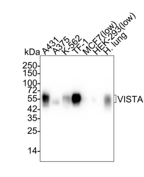
Western blot analysis of VISTA on different lysates with Rabbit anti-VISTA antibody at 1/1,000 dilution. Lane 1: A431 cell lysate (20 µg/Lane), Lane 2: A375 cell lysate (20 µg/Lane), Lane 3: K-562 cell lysate (20 µg/Lane), Lane 4: TF-1 cell lysate (20 µg/Lane), Lane 5: MCF7 cell lysate (low expression) (20 µg/Lane), Lane 6: HEK-293 cell lysate (low expression) (20 µg/Lane), Lane 7: Human lung tissue lysate (30 µg/Lane), Exposure time: 30 seconds; ECL: K1801; 4-20% SDS-PAGE gel. Proteins were transferred to a PVDF membrane and blocked with 5% NFDM/TBST for 1 hour at room temperature. The primary antibody at 1/1,000 dilution was used in 5% NFDM/TBST at 4℃ overnight. Goat Anti-Rabbit IgG - HRP Secondary Antibody at 1/50,000 dilution was used for 1 hour at room temperature.IHC
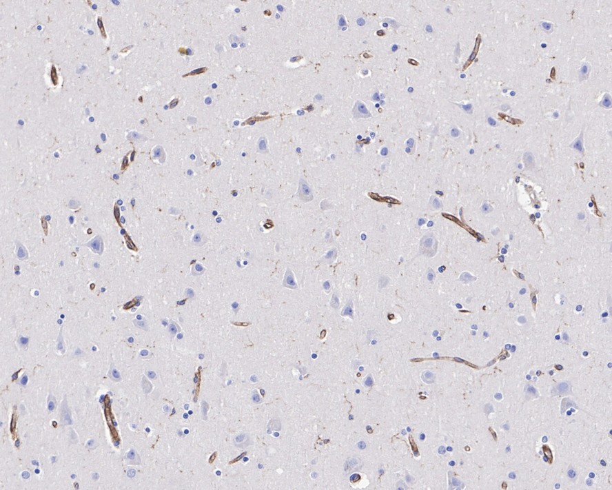
Immunohistochemical analysis of paraffin-embedded human brain tissue with Rabbit anti-VISTA antibody at 1/1,000 dilution. The section was pre-treated using heat mediated antigen retrieval with Tris-EDTA buffer (pH 9.0) for 20 minutes. The tissues were blocked in 1% BSA for 20 minutes at room temperature, washed with ddH2O and PBS, and then probed with the primary antibody at 1/1,000 dilution for 1 hour at room temperature. The detection was performed using an HRP conjugated compact polymer system. DAB was used as the chromogen. Tissues were counterstained with hematoxylin and mounted with DPX.IHC
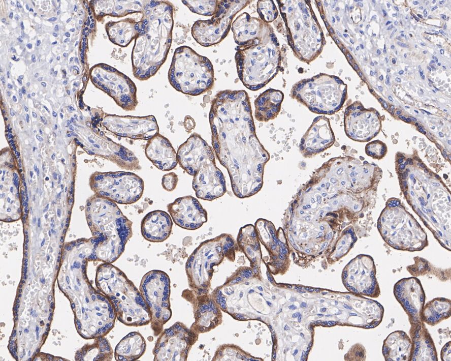
Immunohistochemical analysis of paraffin-embedded human placenta tissue with Rabbit anti-VISTA antibody at 1/1,000 dilution. The section was pre-treated using heat mediated antigen retrieval with Tris-EDTA buffer (pH 9.0) for 20 minutes. The tissues were blocked in 1% BSA for 20 minutes at room temperature, washed with ddH2O and PBS, and then probed with the primary antibody at 1/1,000 dilution for 1 hour at room temperature. The detection was performed using an HRP conjugated compact polymer system. DAB was used as the chromogen. Tissues were counterstained with hematoxylin and mounted with DPX.IHC
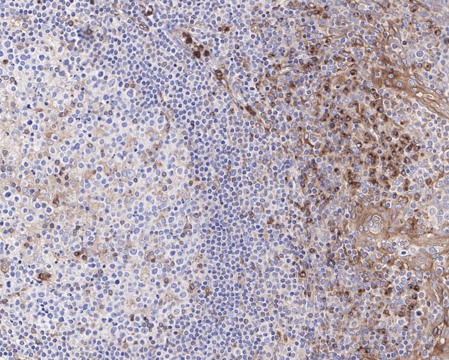
Immunohistochemical analysis of paraffin-embedded human tonsil tissue with Rabbit anti-VISTA antibody at 1/1,000 dilution. The section was pre-treated using heat mediated antigen retrieval with Tris-EDTA buffer (pH 9.0) for 20 minutes. The tissues were blocked in 1% BSA for 20 minutes at room temperature, washed with ddH2O and PBS, and then probed with the primary antibody at 1/1,000 dilution for 1 hour at room temperature. The detection was performed using an HRP conjugated compact polymer system. DAB was used as the chromogen. Tissues were counterstained with hematoxylin and mounted with DPX.IF-P
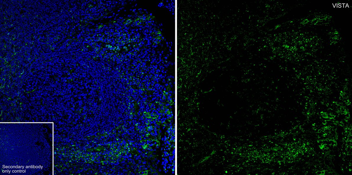
Species: Human, Site: tonsil, Sample: Paraffin-embedded section, Antibody concentration: 1/500.| Product Name | VISTA Recombinant Rabbit Monoclonal Antibody |
|---|---|
| Antibody Type | Primary Antibodies |
| Immunogen | Recombinant protein within human VISTA aa 1-194 / 311. |
| Clonality | monoclonal |
|---|---|
| Isotype | IgG |
| Host Species | Rabbit |
| Tested Applications | IF-PIHCWB |
| WB:1:1000 IHC:1:1000 IF-P:1:500 |
|
| Species Reactivity | Human |
| Concentration | 1mg/ml |
| Purification | Protein A |
| Gene Symbol | VSIR |
|---|---|
| Gene Synonyms | B7H5 GI24 B7-H5 Dies1 PD-1H SISP1 VISTA PP2135 C10orf54 DD1alpha |
| Gene Full Name | V-set immunoregulatory receptor |
| Gene Summary | Enables endopeptidase activator activity; enzyme binding activity; and identical protein binding activity. Involved in several processes, including negative regulation of cytokine production; positive regulation of metabolic process; and regulation of T cell activation. Located in plasma membrane. [provided by Alliance of Genome Resources, Jul 2025] |
| Alternative Names | B7 H5 antibody B7H5 antibody C10orf54 antibody chromosome 10 open reading frame 54 antibody DD1alpha antibody GI24 antibody GI24_HUMAN antibody PD-1H antibody PDCD1 homolog antibody Platelet receptor Gi24 antibody B7 H5 antibody B7H5 antibody C10orf54 antibody chromosome 10 open reading frame 54 antibody DD1alpha antibody GI24 antibody GI24_HUMAN antibody PD-1H antibody PDCD1 homolog antibody Platelet receptor Gi24 antibody PP2135 antibody sisp 1 antibody SISP1 antibody stress induced secreted protein 1 antibody UNQ730/PRO1412 antibody V domain Ig suppressor of T cell activation antibody V set domain containing immunoregulatory receptor antibody V set immunoregulatory receptor antibody V type immunoglobulin domain containing suppressor of T cell activation antibody VISTA antibody |
| Molecular Weight(MW) | 34 kDa(Observed band size:45-60 kDa (Glycosylated)) |
| Cellular Localization | Cell membrane. |
WB
Western blot analysis of VISTA on different lysates with Rabbit anti-VISTA antibody at 1/1,000 dilution. Lane 1: A431 cell lysate (20 µg/Lane), Lane 2: A375 cell lysate (20 µg/Lane), Lane 3: K-562 cell lysate (20 µg/Lane), Lane 4: TF-1 cell lysate (20 µg/Lane), Lane 5: MCF7 cell lysate (low expression) (20 µg/Lane), Lane 6: HEK-293 cell lysate (low expression) (20 µg/Lane), Lane 7: Human lung tissue lysate (30 µg/Lane), Exposure time: 30 seconds; ECL: K1801; 4-20% SDS-PAGE gel. Proteins were transferred to a PVDF membrane and blocked with 5% NFDM/TBST for 1 hour at room temperature. The primary antibody at 1/1,000 dilution was used in 5% NFDM/TBST at 4℃ overnight. Goat Anti-Rabbit IgG - HRP Secondary Antibody at 1/50,000 dilution was used for 1 hour at room temperature.
IHC
Immunohistochemical analysis of paraffin-embedded human brain tissue with Rabbit anti-VISTA antibody at 1/1,000 dilution. The section was pre-treated using heat mediated antigen retrieval with Tris-EDTA buffer (pH 9.0) for 20 minutes. The tissues were blocked in 1% BSA for 20 minutes at room temperature, washed with ddH2O and PBS, and then probed with the primary antibody at 1/1,000 dilution for 1 hour at room temperature. The detection was performed using an HRP conjugated compact polymer system. DAB was used as the chromogen. Tissues were counterstained with hematoxylin and mounted with DPX.
IHC
Immunohistochemical analysis of paraffin-embedded human placenta tissue with Rabbit anti-VISTA antibody at 1/1,000 dilution. The section was pre-treated using heat mediated antigen retrieval with Tris-EDTA buffer (pH 9.0) for 20 minutes. The tissues were blocked in 1% BSA for 20 minutes at room temperature, washed with ddH2O and PBS, and then probed with the primary antibody at 1/1,000 dilution for 1 hour at room temperature. The detection was performed using an HRP conjugated compact polymer system. DAB was used as the chromogen. Tissues were counterstained with hematoxylin and mounted with DPX.
IHC
Immunohistochemical analysis of paraffin-embedded human tonsil tissue with Rabbit anti-VISTA antibody at 1/1,000 dilution. The section was pre-treated using heat mediated antigen retrieval with Tris-EDTA buffer (pH 9.0) for 20 minutes. The tissues were blocked in 1% BSA for 20 minutes at room temperature, washed with ddH2O and PBS, and then probed with the primary antibody at 1/1,000 dilution for 1 hour at room temperature. The detection was performed using an HRP conjugated compact polymer system. DAB was used as the chromogen. Tissues were counterstained with hematoxylin and mounted with DPX.
IF-P
Species: Human, Site: tonsil, Sample: Paraffin-embedded section, Antibody concentration: 1/500.| Application Notes | WB:1:1000 IHC:1:1000 IF-P:1:500 |
|---|
| Form | Liquid |
|---|---|
| Storage Instructions | Store at +4℃ after thawing. Aliquot store at -20℃. Avoid repeated freeze / thaw cycles. |
| Storage Buffer | PBS (pH7.4), 0.1% BSA, 40% Glycerol. Preservative: 0.05% Sodium Azide. |


