IHC
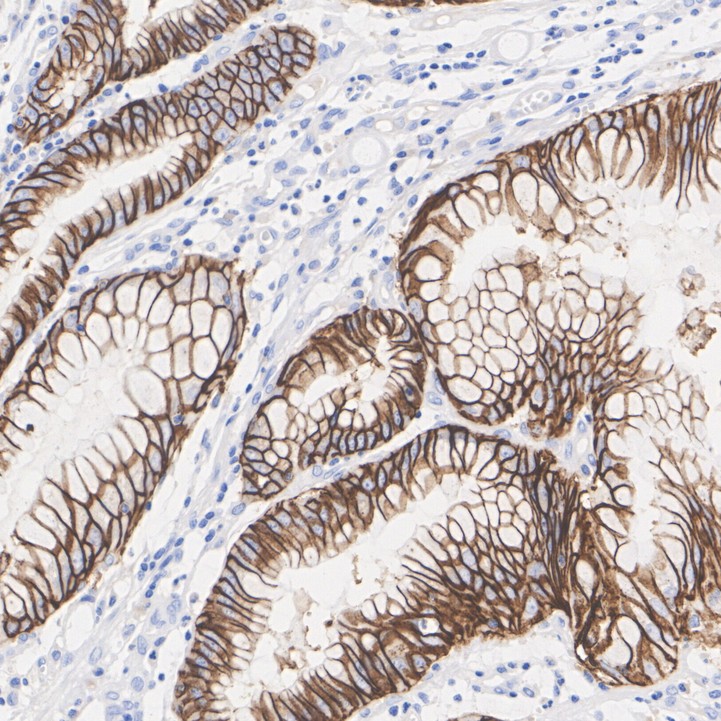
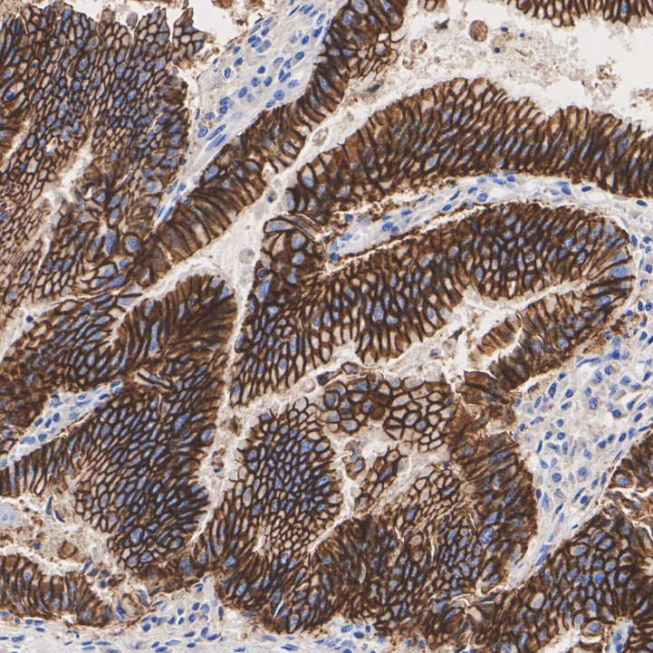
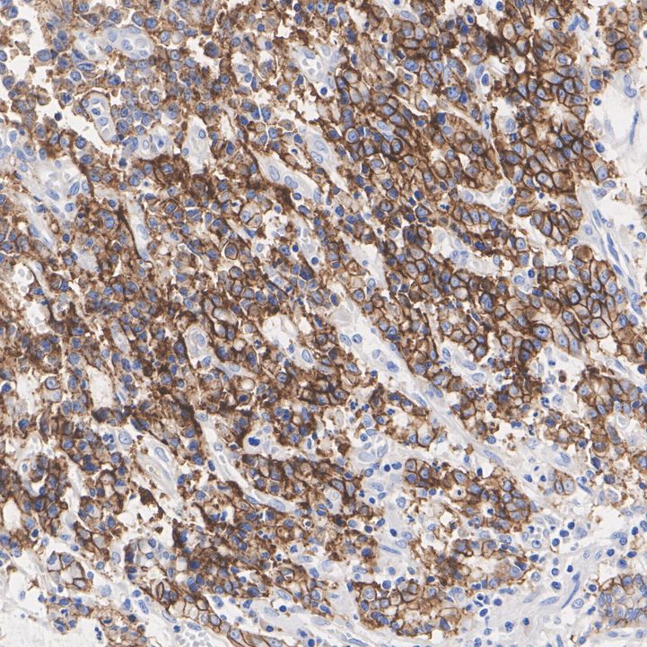
Immunohistochemical analysis of paraffin-embedded human stomach cancer tissue with Mouse anti-Claudin 18.2 antibody at 1/50 dilution. The section was pre-treated using heat mediated antigen retrieval with Tris-EDTA buffer (pH 9.0) for 20 minutes. The tissues were blocked in 1% BSA for 20 minutes at room temperature, washed with ddH2O and PBS, and then probed with the primary antibody at 1/50 dilution for 1 hour at room temperature. The detection was performed using an HRP conjugated compact polymer system. DAB was used as the chromogen. Tissues were counterstained with hematoxylin and mounted with DPX.IHC
Immunohistochemical analysis of paraffin-embedded human stomach cancer tissue with Mouse anti-Claudin 18.2 antibody at 1/50 dilution. The section was pre-treated using heat mediated antigen retrieval with Tris-EDTA buffer (pH 9.0) for 20 minutes. The tissues were blocked in 1% BSA for 20 minutes at room temperature, washed with ddH2O and PBS, and then probed with the primary antibody at 1/50 dilution for 1 hour at room temperature. The detection was performed using an HRP conjugated compact polymer system. DAB was used as the chromogen. Tissues were counterstained with hematoxylin and mounted with DPX.IHC
Immunohistochemical analysis of paraffin-embedded human stomach cancer tissue with Mouse anti-Claudin 18.2 antibody at 1/50 dilution. The section was pre-treated using heat mediated antigen retrieval with Tris-EDTA buffer (pH 9.0) for 20 minutes. The tissues were blocked in 1% BSA for 20 minutes at room temperature, washed with ddH2O and PBS, and then probed with the primary antibody at 1/50 dilution for 1 hour at room temperature. The detection was performed using an HRP conjugated compact polymer system. DAB was used as the chromogen. Tissues were counterstained with hematoxylin and mounted with DPX.| Product Name | Claudin 18.2 Recombinant Mouse Monoclonal Antibody |
|---|---|
| Antibody Type | Primary Antibodies |
| Immunogen | Recombinant protein. |
| Clonality | monoclonal |
|---|---|
| Isotype | IgG |
| Host Species | Mouse |
| Tested Applications | IHC |
| IHC:1:50-1:200 |
|
| Species Reactivity | Human |
| Concentration | 1mg/ml |
| Purification | Protein A |
| Gene Symbol | CLDN18 |
|---|---|
| Gene Synonyms | SFTA5 SFTPJ |
| Gene Full Name | claudin 18 |
| Gene Summary | This gene encodes a member of the claudin family. Claudins are integral membrane proteins and components of tight junction strands. Tight junction strands serve as a physical barrier to prevent solutes and water from passing freely through the paracellular space between epithelial or endothelial cell sheets, and also play critical roles in maintaining cell polarity and signal transductions. This gene is upregulated in patients with ulcerative colitis and highly overexpressed in infiltrating ductal adenocarcinomas. PKC/MAPK/AP-1 (protein kinase C/mitogen-activated protein kinase/activator protein-1) dependent pathway regulates the expression of this gene in gastric cells. Alternatively spliced transcript variants encoding different isoforms have been identified. [provided by RefSeq, Jun 2010] |
| Alternative Names | Claudin 18 antibody Claudin-18 antibody CLD18_HUMAN antibody CLDN18 antibody SFTA5 antibody SFTPJ antibody CLDN18.2 |
| Molecular Weight(MW) | 28kDa |
| Function | CLDN-18 has two isoforms, CLDN-18.1 and CLDN-18.2, that are expressed mainly in normal lung and gastric tissues, respectively. CLDN-18 is expressed in many neoplasias, such as gastric and gastroesophageal junction (G/GEJ) cancer, pancreatic cancer, head and neck cancer and non-small cell lung cancer. Different therapeutic drugs targeting CLDN-18.2 for cancer immunotherapy have now been developed and IHC for CLDN-18.2 to be used for the selection of patients to be offered anti-CLDN18.2 treatment. The distinction and scoring criteria separating CLDN-18.2 positive cancers versus negative cancers depend on tumour type and therapeutic agent. For G/GEJ cancer a moderate to strong (2+/3+) positive membrane staining in ≥75% of tumor cells should be considered as the standard cut-off for a CLDN18.2-positive tumor to be treated with zolbetuximab (VYLOY™, Astellas). CLDN-18.2 has a very limited expression in normal tissues and is exclusively expressed in the mucosal membrane of differentiated gastric epithelial cells complicating the design of guidelines for composition of control material for CLDN-18.2 IHC. The gastric epithelial cells will typically show a moderate to strong membranous staining reaction and thus mainly applicable to confirm “right IHC assay” being applied. No staining reaction should be seen in e.g. smooth muscle cells, lymphocytes and endothelial cells enabling these structures to be used as negative tissue controls. Gastric intestinal metaplasia is reported and shown to have a reduced CLDN-18.2 expression and potential to be used as immunohistochemical critical assay performance control (iCAPC) with emphasis on low level demonstration of CLDN-18.2. Gastric intestinal metaplastic epithelial cells (either as external tissue control or internally in patient sample) will typically show a weak to moderate staining reaction. At present internal NordiQC testing has revealed that the presence of gastric intestinal metaplasia is very focal and heterogeneously observed compromising the use for iCAPC in external tissue controls. |
| Cellular Localization | Cell junction, tight junction, Lateral cell membrane. |
IHC
Immunohistochemical analysis of paraffin-embedded human stomach cancer tissue with Mouse anti-Claudin 18.2 antibody at 1/50 dilution. The section was pre-treated using heat mediated antigen retrieval with Tris-EDTA buffer (pH 9.0) for 20 minutes. The tissues were blocked in 1% BSA for 20 minutes at room temperature, washed with ddH2O and PBS, and then probed with the primary antibody at 1/50 dilution for 1 hour at room temperature. The detection was performed using an HRP conjugated compact polymer system. DAB was used as the chromogen. Tissues were counterstained with hematoxylin and mounted with DPX.
IHC
Immunohistochemical analysis of paraffin-embedded human stomach cancer tissue with Mouse anti-Claudin 18.2 antibody at 1/50 dilution. The section was pre-treated using heat mediated antigen retrieval with Tris-EDTA buffer (pH 9.0) for 20 minutes. The tissues were blocked in 1% BSA for 20 minutes at room temperature, washed with ddH2O and PBS, and then probed with the primary antibody at 1/50 dilution for 1 hour at room temperature. The detection was performed using an HRP conjugated compact polymer system. DAB was used as the chromogen. Tissues were counterstained with hematoxylin and mounted with DPX.
IHC
Immunohistochemical analysis of paraffin-embedded human stomach cancer tissue with Mouse anti-Claudin 18.2 antibody at 1/50 dilution. The section was pre-treated using heat mediated antigen retrieval with Tris-EDTA buffer (pH 9.0) for 20 minutes. The tissues were blocked in 1% BSA for 20 minutes at room temperature, washed with ddH2O and PBS, and then probed with the primary antibody at 1/50 dilution for 1 hour at room temperature. The detection was performed using an HRP conjugated compact polymer system. DAB was used as the chromogen. Tissues were counterstained with hematoxylin and mounted with DPX.| Application Notes | IHC:1:50-1:200 |
|---|
| Form | Liquid |
|---|---|
| Storage Instructions | Store at +4℃ after thawing. Aliquot store at -20℃. Avoid repeated freeze / thaw cycles. |
| Storage Buffer | PBS (pH7.4), 0.1% BSA. Preservative: 0.05% Sodium Azide. |


