WB
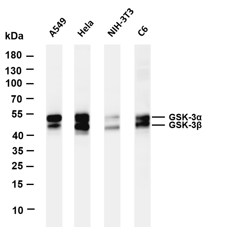
Various whole cell lysates were separated by 4-20% SDS-PAGE, and the membrane was blotted with anti-GSK3 α/β antibody. The HRP-conjugated Goat anti-Rabbit IgG(H + L) antibody was used to detect the antibody. Lane 1: A549 Lane 2: Hela Lane 3: NIH-3T3 Lane 4: C6 Predicted band size: 51,47kDa Observed band size: 51,47kDaWB
Various whole cell lysates were separated by 4-20% SDS-PAGE, and the membrane was blotted with anti-GSK3 α/β antibody. The HRP-conjugated Goat anti-Rabbit IgG(H + L) antibody was used to detect the antibody. Lane 1: A549 Lane 2: Hela Lane 3: NIH-3T3 Lane 4: C6 Predicted band size: 51,47kDa Observed band size: 51,47kDaWB
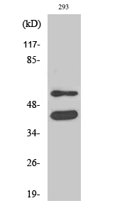
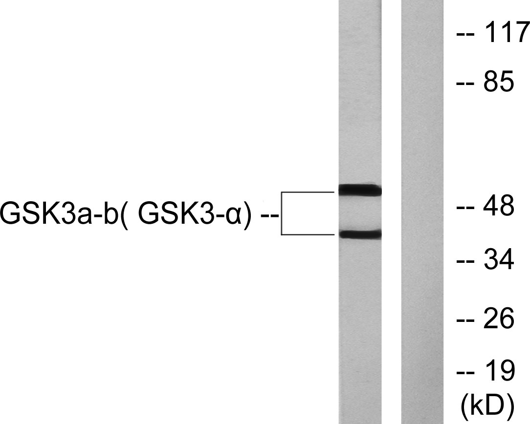
Western blot analysis of lysates from 293 cells, treated with TNF-a 20ng/ml 30', using GSK3 alpha/beta Antibody. The lane on the right is blocked with the synthesized peptide.IHC
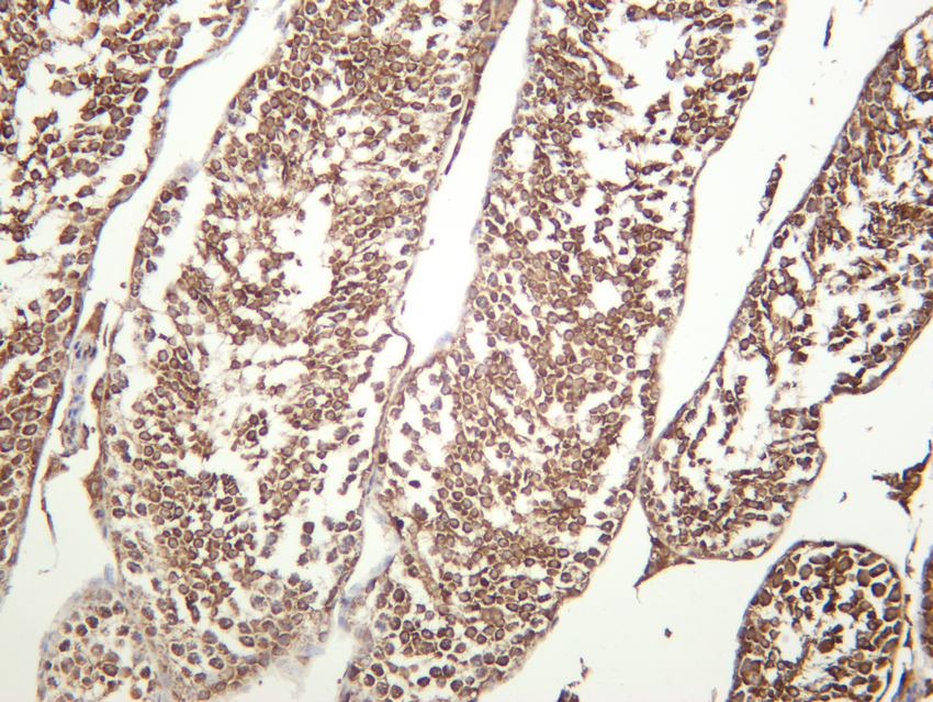
Mouse testis was stained with anti-GSK3 α/β rabbit antibodyIHC
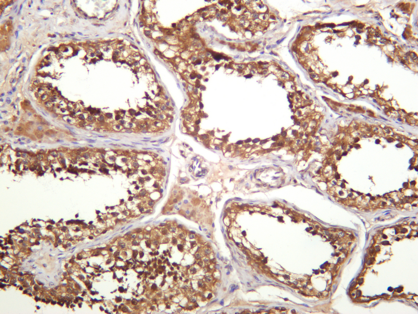
Human testis was stained with anti-GSK3 α/β rabbit antibodyIF/ICC
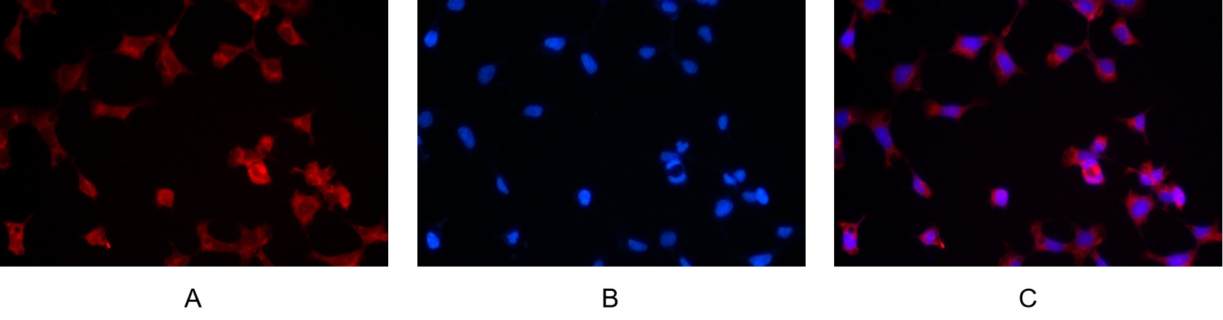
Immunofluorescence analysis of HEK293. Picture A: GSK3 α/β antibody (red). Picture B: DAPI (blue). Picture C: Merge of A+B| Product Name | GSK3α/β Rabbit mAb |
|---|---|
| Antibody Type | Primary Antibodies |
| Immunogen | Glycogen synthase kinase-3 alpha/beta |
| Clonality | monoclonal |
|---|---|
| Isotype | IgG |
| Host Species | Rabbit |
| Tested Applications | ELISAICC/IFIHCIPWB |
| WB:1:2000-1:10000 IHC: 1:400-1:800 IF: 1:200-1:1000 ELISA: 1:5000-1:20000 IP:1:50-1:200 Note:For IHC,wesuggest antigen retrieval with TE buffer pH 9.0ICC/IF:1:50-1:200 |
|
| Species Reactivity | HumanMouseRat |
| Concentration | 1mg/ml |
| Purification | Protein A |
| Gene Symbol | GSK3A |
|---|---|
| Gene Full Name | glycogen synthase kinase 3 alpha |
| Gene Summary | This gene encodes a multifunctional Ser/Thr protein kinase that is implicated in the control of several regulatory proteins including glycogen synthase, and transcription factors, such as JUN. It also plays a role in the WNT and PI3K signaling pathways, as well as regulates the production of beta-amyloid peptides associated with Alzheimer's disease. [provided by RefSeq, Oct 2011] |
| Alternative Names | GSK3A Glycogen synthase kinase-3 alpha GSK-3 alpha Serine/threonine-protein kinase GSK3A GSK3B Glycogen synthase kinase-3 beta GSK-3 beta Serine/threonine-protein kinase GSK3B |
| Molecular Weight(MW) | 51kD,47kD |
| Cellular Localization | Cytoplasm |
WB
Various whole cell lysates were separated by 4-20% SDS-PAGE, and the membrane was blotted with anti-GSK3 α/β antibody. The HRP-conjugated Goat anti-Rabbit IgG(H + L) antibody was used to detect the antibody. Lane 1: A549 Lane 2: Hela Lane 3: NIH-3T3 Lane 4: C6 Predicted band size: 51,47kDa Observed band size: 51,47kDa
WB
Various whole cell lysates were separated by 4-20% SDS-PAGE, and the membrane was blotted with anti-GSK3 α/β antibody. The HRP-conjugated Goat anti-Rabbit IgG(H + L) antibody was used to detect the antibody. Lane 1: A549 Lane 2: Hela Lane 3: NIH-3T3 Lane 4: C6 Predicted band size: 51,47kDa Observed band size: 51,47kDa
WB
Western blot analysis of lysates from 293 cells, treated with TNF-a 20ng/ml 30', using GSK3 alpha/beta Antibody. The lane on the right is blocked with the synthesized peptide.
IHC
Mouse testis was stained with anti-GSK3 α/β rabbit antibody
IHC
Human testis was stained with anti-GSK3 α/β rabbit antibody
IF/ICC
Immunofluorescence analysis of HEK293. Picture A: GSK3 α/β antibody (red). Picture B: DAPI (blue). Picture C: Merge of A+B| Application Notes | WB:1:2000-1:10000 IHC: 1:400-1:800 IF: 1:200-1:1000 ELISA: 1:5000-1:20000 IP:1:50-1:200 Note:For IHC,wesuggest antigen retrieval with TE buffer pH 9.0ICC/IF:1:50-1:200 |
|---|
| Form | Liquid |
|---|---|
| Storage Instructions | -15°C to -25°C/1 year(Do not lower than -25°C) |
| Storage Buffer | PBS, 50% glycerol, 0.05% Proclin 300, 0.05%BSA. |


