WB
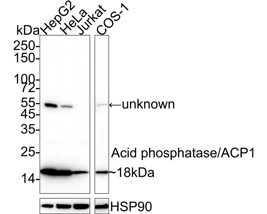
Western blot analysis of Acid phosphatase/ACP1 on different lysates with Rabbit anti-Acid phosphatase/ACP1 antibody at 1/2,000 dilution. Lane 1: HepG2 cell lysate, Lane 2: HeLa cell lysate, Lane 3: Jurkat cell lysate, Lane 4: COS-1 cell lysate, Lysates/proteins at 20 µg/Lane. Exposure time: 1 minute 33 seconds; 4-20% SDS-PAGE gel. Proteins were transferred to a PVDF membrane and blocked with 5% NFDM/TBST for 1 hour at room temperature. The primary antibody at 1/2,000 dilution was used in 5% NFDM/TBST at 4℃ overnight. Goat Anti-Rabbit IgG - HRP Secondary Antibody at 1/50,000 dilution was used for 1 hour at room temperature.IHC
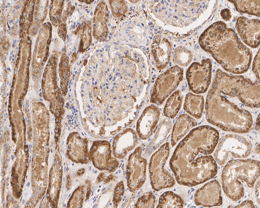
Immunohistochemical analysis of paraffin-embedded human kidney tissue with Rabbit anti-Acid phosphatase/ACP1 antibody at 1/1,000 dilution. The section was pre-treated using heat mediated antigen retrieval with Tris-EDTA buffer (pH 9.0) for 20 minutes. The tissues were blocked in 1% BSA for 20 minutes at room temperature, washed with ddH2O and PBS, and then probed with the primary antibody at 1/1,000 dilution for 1 hour at room temperature. The detection was performed using an HRP conjugated compact polymer system. DAB was used as the chromogen. Tissues were counterstained with hematoxylin and mounted with DPX.IHC
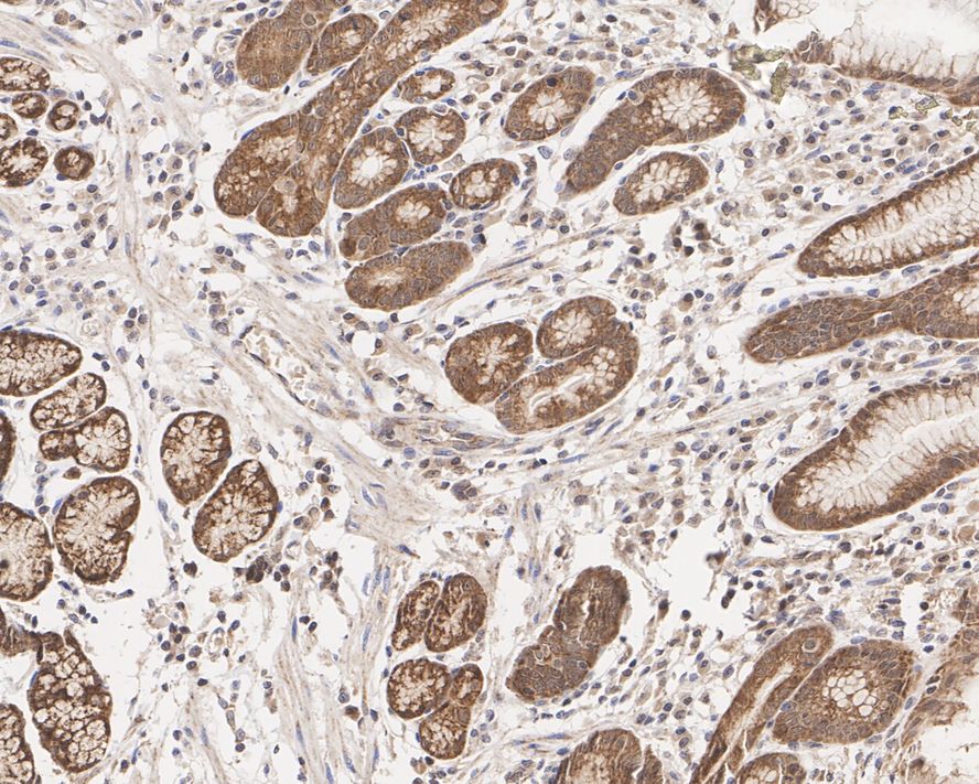
Immunohistochemical analysis of paraffin-embedded human stomach tissue with Rabbit anti-Acid phosphatase/ACP1 antibody at 1/1,000 dilution. The section was pre-treated using heat mediated antigen retrieval with Tris-EDTA buffer (pH 9.0) for 20 minutes. The tissues were blocked in 1% BSA for 20 minutes at room temperature, washed with ddH2O and PBS, and then probed with the primary antibody at 1/1,000 dilution for 1 hour at room temperature. The detection was performed using an HRP conjugated compact polymer system. DAB was used as the chromogen. Tissues were counterstained with hematoxylin and mounted with DPX.IHC
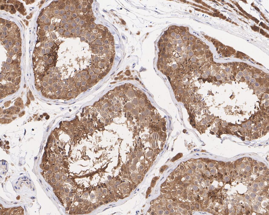
Immunohistochemical analysis of paraffin-embedded human testis tissue with Rabbit anti-Acid phosphatase/ACP1 antibody at 1/1,000 dilution. The section was pre-treated using heat mediated antigen retrieval with Tris-EDTA buffer (pH 9.0) for 20 minutes. The tissues were blocked in 1% BSA for 20 minutes at room temperature, washed with ddH2O and PBS, and then probed with the primary antibody at 1/1,000 dilution for 1 hour at room temperature. The detection was performed using an HRP conjugated compact polymer system. DAB was used as the chromogen. Tissues were counterstained with hematoxylin and mounted with DPX.| Product Name | Acid phosphatase/ACP1 Recombinant Rabbit Monoclonal Antibody |
|---|---|
| Antibody Type | Primary Antibodies |
| Immunogen | Recombinant protein within human ACP1 aa 1-158 / 158. |
| Clonality | monoclonal |
|---|---|
| Isotype | IgG |
| Host Species | Rabbit |
| Tested Applications | IHCWB |
| WB:1:2000 IHC:1:1000 |
|
| Species Reactivity | HumanMonkey |
| Concentration | 1mg/ml |
| Purification | Protein A |
| Gene Symbol | ACP1 |
|---|---|
| Gene Synonyms | HAAP LMWPTP LMW-PTP |
| Gene Full Name | acid phosphatase 1 |
| Gene Summary | The product of this gene belongs to the phosphotyrosine protein phosphatase family of proteins. It functions as an acid phosphatase and a protein tyrosine phosphatase by hydrolyzing protein tyrosine phosphate to protein tyrosine and orthophosphate. This enzyme also hydrolyzes orthophosphoric monoesters to alcohol and orthophosphate. This gene is genetically polymorphic, and three common alleles segregating at the corresponding locus give rise to six phenotypes. Each allele appears to encode at least two electrophoretically different isozymes, Bf and Bs, which are produced in allele-specific ratios. Multiple alternatively spliced transcript variants encoding distinct isoforms have been identified for this gene. [provided by RefSeq, Aug 2008] |
| Alternative Names | Acid phosphatase 1 soluble antibody Acid phosphatase of erythrocyte antibody ACP1 antibody Adipocyte acid phosphatase antibody Cytoplasmic phosphotyrosyl protein phosphatase antibody HAAP antibody LMW-PTP antibody LMW-PTPase antibody Low molecular weight cytosolic acid phosphatase antibody Low molecular weight phosphotyrosine protein phosphatase antibody Acid phosphatase 1 soluble antibody Acid phosphatase of erythrocyte antibody ACP1 antibody Adipocyte acid phosphatase antibody Cytoplasmic phosphotyrosyl protein phosphatase antibody HAAP antibody LMW-PTP antibody LMW-PTPase antibody Low molecular weight cytosolic acid phosphatase antibody Low molecular weight phosphotyrosine protein phosphatase antibody PAP1 antibody PAP2 antibody phosphatase acid of erythrocyte antibody PPAC_HUMAN antibody Protein tyrosine phosphatase antibody PTPase antibody Purple acid phosphatase antibody Red cell acid phosphatase 1 antibody testicular secretory protein Li 37 antibody |
| Molecular Weight(MW) | 18kDa |
| Cellular Localization | Cytoplasm. |
WB
Western blot analysis of Acid phosphatase/ACP1 on different lysates with Rabbit anti-Acid phosphatase/ACP1 antibody at 1/2,000 dilution. Lane 1: HepG2 cell lysate, Lane 2: HeLa cell lysate, Lane 3: Jurkat cell lysate, Lane 4: COS-1 cell lysate, Lysates/proteins at 20 µg/Lane. Exposure time: 1 minute 33 seconds; 4-20% SDS-PAGE gel. Proteins were transferred to a PVDF membrane and blocked with 5% NFDM/TBST for 1 hour at room temperature. The primary antibody at 1/2,000 dilution was used in 5% NFDM/TBST at 4℃ overnight. Goat Anti-Rabbit IgG - HRP Secondary Antibody at 1/50,000 dilution was used for 1 hour at room temperature.
IHC
Immunohistochemical analysis of paraffin-embedded human kidney tissue with Rabbit anti-Acid phosphatase/ACP1 antibody at 1/1,000 dilution. The section was pre-treated using heat mediated antigen retrieval with Tris-EDTA buffer (pH 9.0) for 20 minutes. The tissues were blocked in 1% BSA for 20 minutes at room temperature, washed with ddH2O and PBS, and then probed with the primary antibody at 1/1,000 dilution for 1 hour at room temperature. The detection was performed using an HRP conjugated compact polymer system. DAB was used as the chromogen. Tissues were counterstained with hematoxylin and mounted with DPX.
IHC
Immunohistochemical analysis of paraffin-embedded human stomach tissue with Rabbit anti-Acid phosphatase/ACP1 antibody at 1/1,000 dilution. The section was pre-treated using heat mediated antigen retrieval with Tris-EDTA buffer (pH 9.0) for 20 minutes. The tissues were blocked in 1% BSA for 20 minutes at room temperature, washed with ddH2O and PBS, and then probed with the primary antibody at 1/1,000 dilution for 1 hour at room temperature. The detection was performed using an HRP conjugated compact polymer system. DAB was used as the chromogen. Tissues were counterstained with hematoxylin and mounted with DPX.
IHC
Immunohistochemical analysis of paraffin-embedded human testis tissue with Rabbit anti-Acid phosphatase/ACP1 antibody at 1/1,000 dilution. The section was pre-treated using heat mediated antigen retrieval with Tris-EDTA buffer (pH 9.0) for 20 minutes. The tissues were blocked in 1% BSA for 20 minutes at room temperature, washed with ddH2O and PBS, and then probed with the primary antibody at 1/1,000 dilution for 1 hour at room temperature. The detection was performed using an HRP conjugated compact polymer system. DAB was used as the chromogen. Tissues were counterstained with hematoxylin and mounted with DPX.| Application Notes | WB:1:2000 IHC:1:1000 |
|---|
| Form | Liquid |
|---|---|
| Storage Instructions | Store at +4℃ after thawing. Aliquot store at -20℃. Avoid repeated freeze / thaw cycles. |
| Storage Buffer | PBS (pH7.4), 0.1% BSA, 40% Glycerol. Preservative: 0.05% Sodium Azide. |


