WB
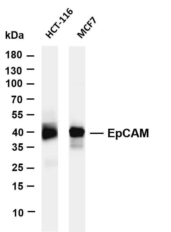
Various whole cell lysates were separated by 4-20% SDS-PAGE, and the membrane was blotted with anti-EpCAM antibody. The HRP-conjugated Goat anti-Rabbit IgG(H + L) antibody was used to detect the antibody. Lane 1: HCT-116, Lane 2: MCF-7.IHC
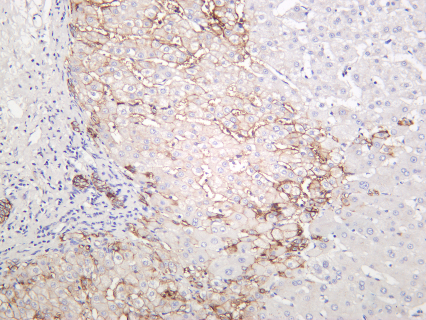
Human liver was stained with anti-EpCAM rabbit antibody.IHC
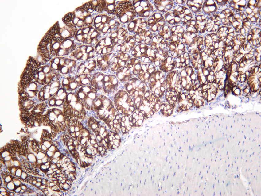
Mouse colon was stained with anti-EpCAM rabbit antibody.IHC
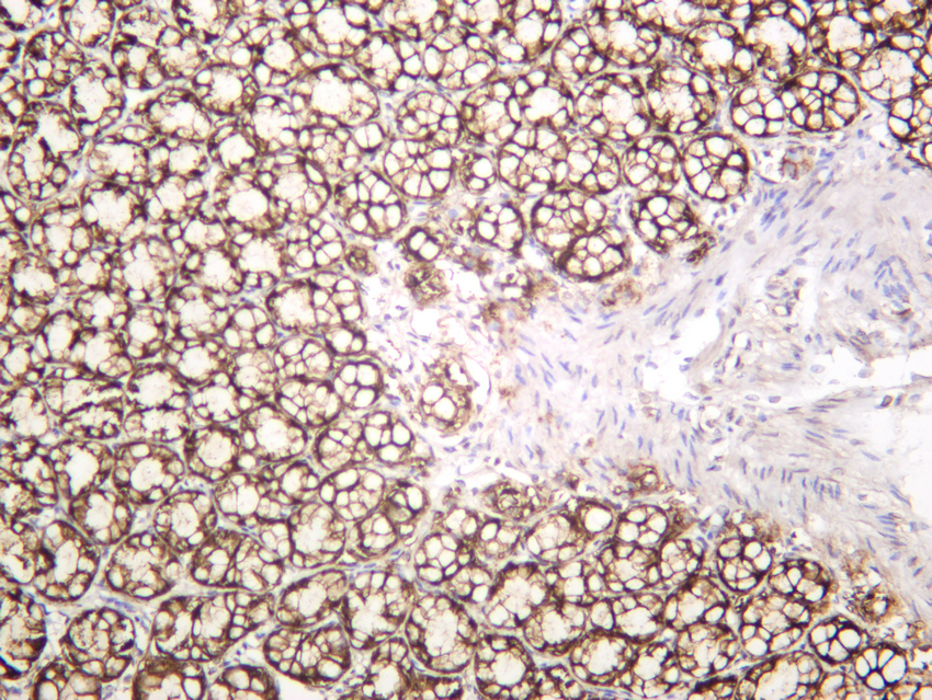
Rat colon was stained with anti-EpCAM rabbit antibody.ICC/IF
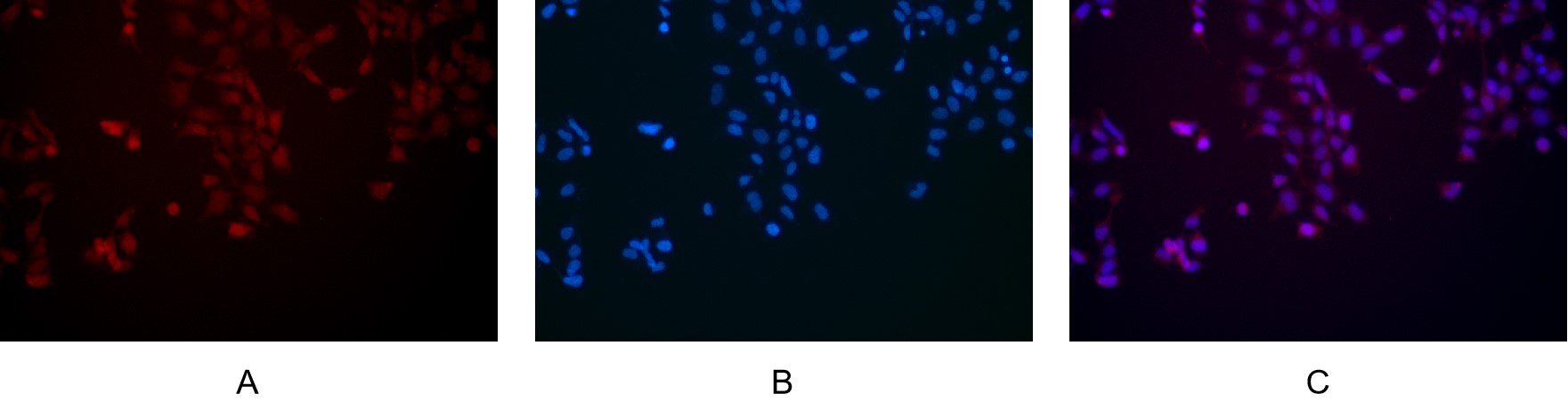
Immunofluorescence analysis of HEK293. Picture A: EpCAM antibody (red). Picture B: DAPI (blue). Picture C: Merge of A+B.IF-P
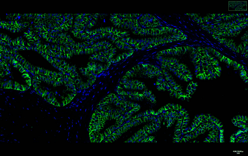
Rat prostate was stained with anti-EpCAM Rabbit antibody.IF-P
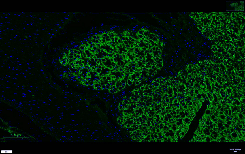
Mouse colon was stained with anti-EpCAM rabbit antibody.| Product Name | EpCAM Rabbit mAb |
|---|---|
| Antibody Type | Primary Antibodies |
| Clonality | monoclonal |
|---|---|
| Isotype | IgG |
| Host Species | Rabbit |
| Tested Applications | ICC/IFIF-PIHCWB |
| WB:1:1000-1:5000 IHC:1:200-1:1000 ICC/IF:1:200-1:1000 IF-P:1:200-1:1000 |
|
| Species Reactivity | HumanMouseRat |
| Concentration | 1mg/ml |
| Purification | Protein A |
| Gene Symbol | EPCAM |
|---|---|
| Gene Synonyms | ESA KSA M4S1 MK-1 DIAR5 EGP-2 EGP40 KS1/4 MIC18 TROP1 BerEp4 EGP314 HNPCC8 LYNCH8 MOC-31 Ber-Ep4 TACSTD1 |
| Gene Full Name | epithelial cell adhesion molecule |
| Gene Summary | This gene encodes a carcinoma-associated antigen and is a member of a family that includes at least two type I membrane proteins. This antigen is expressed on most normal epithelial cells and gastrointestinal carcinomas and functions as a homotypic calcium-independent cell adhesion molecule. The antigen is being used as a target for immunotherapy treatment of human carcinomas. Mutations in this gene result in congenital tufting enteropathy. [provided by RefSeq, Dec 2008] |
| Alternative Names | EPCAM GA733-2 M1S2 M4S1 MIC18 TACSTD1 TROP1 Epithelial cell adhesion molecule Ep-CAM Adenocarcinoma-associated antigen Cell surface glycoprotein Trop-1 Epithelial cell surface antigen Epithelial glycoprotein EGP Epithelial gly |
| Molecular Weight(MW) | 35kDa(Observed MW 40kDa) |
| Cellular Localization | Membrane. |
WB
Various whole cell lysates were separated by 4-20% SDS-PAGE, and the membrane was blotted with anti-EpCAM antibody. The HRP-conjugated Goat anti-Rabbit IgG(H + L) antibody was used to detect the antibody. Lane 1: HCT-116, Lane 2: MCF-7.
IHC
Human liver was stained with anti-EpCAM rabbit antibody.
IHC
Mouse colon was stained with anti-EpCAM rabbit antibody.
IHC
Rat colon was stained with anti-EpCAM rabbit antibody.
ICC/IF
Immunofluorescence analysis of HEK293. Picture A: EpCAM antibody (red). Picture B: DAPI (blue). Picture C: Merge of A+B.
IF-P
Rat prostate was stained with anti-EpCAM Rabbit antibody.
IF-P
Mouse colon was stained with anti-EpCAM rabbit antibody.| Application Notes | WB:1:1000-1:5000 IHC:1:200-1:1000 ICC/IF:1:200-1:1000 IF-P:1:200-1:1000 |
|---|
| Form | Liquid |
|---|---|
| Storage Instructions | -15°C to -25°C/1 year(Do not lower than -25°C) |
| Storage Buffer | PBS, 50% glycerol, 0.05% Proclin 300, 0.05%BSA. |


