WB
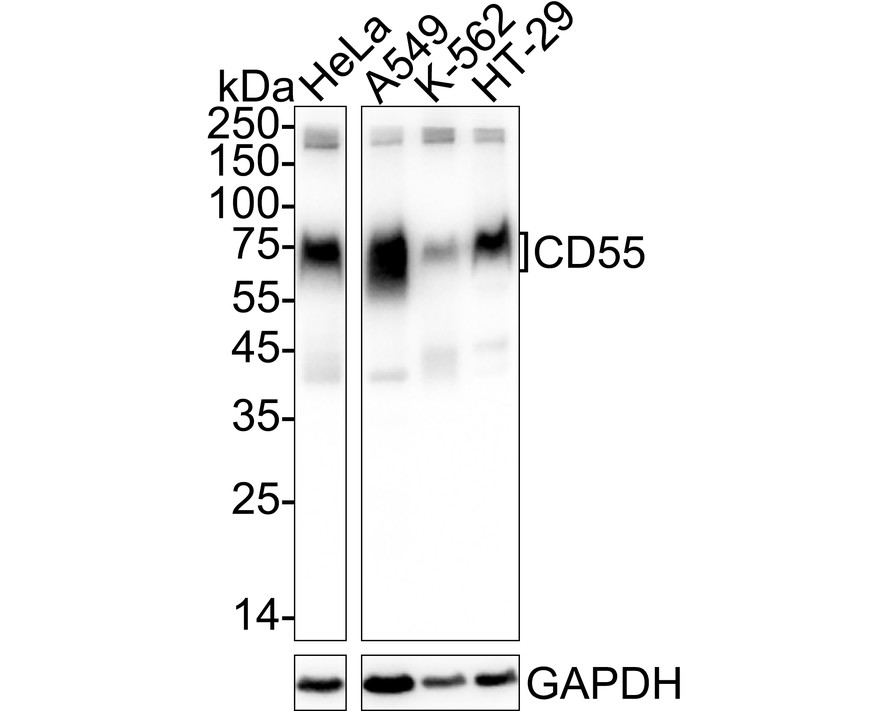
Western blot analysis of CD55 on different lysates with Rabbit anti-CD55 antibody at 1/5,000 dilution. Lane 1: HeLa cell lysate (20 µg/Lane), Lane 2: A549 cell lysate (20 µg/Lane), Lane 3: K-562 cell lysate (20 µg/Lane), Lane 4: HT-29 cell lysate (20 µg/Lane), Exposure time: 2 seconds; 4-20% SDS-PAGE gel. Proteins were transferred to a PVDF membrane and blocked with 5% NFDM/TBST for 1 hour at room temperature. The primary antibody at 1/5,000 dilution was used in 5% NFDM/TBST at 4℃ overnight. Goat Anti-Rabbit IgG - HRP Secondary Antibody at 1/50,000 dilution was used for 1 hour at room temperature.IHC
Western blot analysis of CD55 on different lysates with Rabbit anti-CD55 antibody at 1/5,000 dilution. Lane 1: HeLa cell lysate (20 µg/Lane), Lane 2: A549 cell lysate (20 µg/Lane), Lane 3: K-562 cell lysate (20 µg/Lane), Lane 4: HT-29 cell lysate (20 µg/Lane), Exposure time: 2 seconds; 4-20% SDS-PAGE gel. Proteins were transferred to a PVDF membrane and blocked with 5% NFDM/TBST for 1 hour at room temperature. The primary antibody at 1/5,000 dilution was used in 5% NFDM/TBST at 4℃ overnight. Goat Anti-Rabbit IgG - HRP Secondary Antibody at 1/50,000 dilution was used for 1 hour at room temperature.IHC
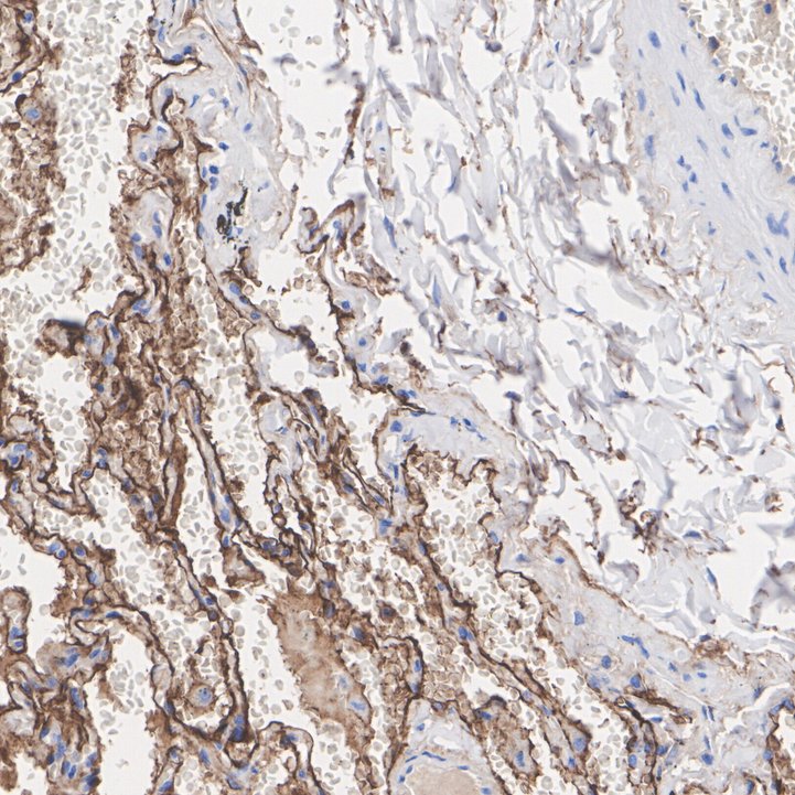
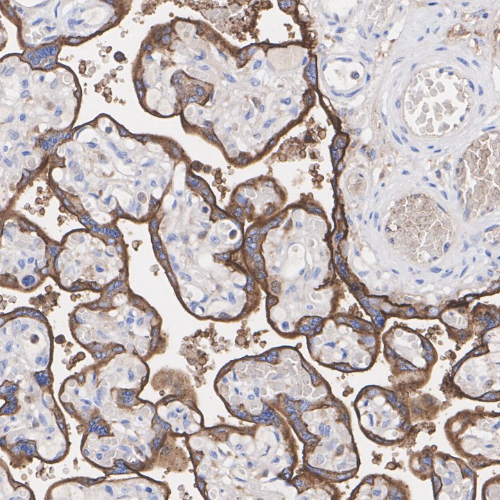
Immunohistochemical analysis of paraffin-embedded human placenta tissue with Rabbit anti-CD55 antibody at 1/200 dilution. The section was pre-treated using heat mediated antigen retrieval with Tris-EDTA buffer (pH 9.0) for 20 minutes. The tissues were blocked in 1% BSA for 20 minutes at room temperature, washed with ddH2O and PBS, and then probed with the primary antibody at 1/200 dilution for 1 hour at room temperature. The detection was performed using an HRP conjugated compact polymer system. DAB was used as the chromogen. Tissues were counterstained with hematoxylin and mounted with DPX.| Product Name | CD55 Recombinant Rabbit Monoclonal Antibody |
|---|---|
| Antibody Type | Primary Antibodies |
| Immunogen | Synthetic peptide within human CD55 aa 101-150 / 381. |
| Clonality | Monoclonal |
|---|---|
| Isotype | IgG |
| Host Species | Rabbit |
| Tested Applications | IHCWB |
| WB:1:5000 IHC:1:200 |
|
| Species Reactivity | Human |
| Concentration | 1mg/ml |
| Purification | Protein A |
| Gene Symbol | CD55 |
|---|---|
| Gene Synonyms | CR TC DAF CROM CHAPLE |
| Gene Full Name | CD55 molecule (Cromer blood group) |
| Gene Summary | This gene encodes a glycoprotein involved in the regulation of the complement cascade. Binding of the encoded protein to complement proteins accelerates their decay, thereby disrupting the cascade and preventing damage to host cells. Antigens present on this protein constitute the Cromer blood group system (CROM). Alternative splicing results in multiple transcript variants. The predominant transcript variant encodes a membrane-bound protein, but alternatively spliced transcripts may produce soluble proteins. [provided by RefSeq, Jul 2014] |
| Alternative Names | CD 55 antibody CD55 antibody CD55 antigen antibody CD55 Cromer blood group system antibody CD55 molecule (Cromer blood group) antibody CD55 molecule antibody CD55 molecule decay accelerating factor for complement (Cromer blood group) antibody Cd55a antibody Complement decay accelerating factor antibody Complement decay-accelerating factor antibody CD 55 antibody CD55 antibody CD55 antigen antibody CD55 Cromer blood group system antibody CD55 molecule (Cromer blood group) antibody CD55 molecule antibody CD55 molecule decay accelerating factor for complement (Cromer blood group) antibody Cd55a antibody Complement decay accelerating factor antibody Complement decay-accelerating factor antibody Complement decay-accelerating factor GPI-anchored antibody CR antibody CROM antibody Cromer Blood Group antigen antibody Cromer blood group system antibody DAF antibody Daf-GPI antibody DAF_HUMAN antibody Daf1 antibody Dcay accelerating factor for complement (CD55 Cromer blood group system) antibody Decay accelarating factor 1 isoform CRA_a antibody Decay accelerating factor (GPI-form) antibody Decay Accelerating Factor for Complement antibody Decay accelerating factor GPI-form antibody Decay accelerating factor soluble-form antibody GPI-DAF antibody TC antibody |
| Molecular Weight(MW) | 41kDa(Observed band size: 70-75kDa) |
| Cellular Localization | Cell membrane. |
WB
Western blot analysis of CD55 on different lysates with Rabbit anti-CD55 antibody at 1/5,000 dilution. Lane 1: HeLa cell lysate (20 µg/Lane), Lane 2: A549 cell lysate (20 µg/Lane), Lane 3: K-562 cell lysate (20 µg/Lane), Lane 4: HT-29 cell lysate (20 µg/Lane), Exposure time: 2 seconds; 4-20% SDS-PAGE gel. Proteins were transferred to a PVDF membrane and blocked with 5% NFDM/TBST for 1 hour at room temperature. The primary antibody at 1/5,000 dilution was used in 5% NFDM/TBST at 4℃ overnight. Goat Anti-Rabbit IgG - HRP Secondary Antibody at 1/50,000 dilution was used for 1 hour at room temperature.
IHC
Western blot analysis of CD55 on different lysates with Rabbit anti-CD55 antibody at 1/5,000 dilution. Lane 1: HeLa cell lysate (20 µg/Lane), Lane 2: A549 cell lysate (20 µg/Lane), Lane 3: K-562 cell lysate (20 µg/Lane), Lane 4: HT-29 cell lysate (20 µg/Lane), Exposure time: 2 seconds; 4-20% SDS-PAGE gel. Proteins were transferred to a PVDF membrane and blocked with 5% NFDM/TBST for 1 hour at room temperature. The primary antibody at 1/5,000 dilution was used in 5% NFDM/TBST at 4℃ overnight. Goat Anti-Rabbit IgG - HRP Secondary Antibody at 1/50,000 dilution was used for 1 hour at room temperature.
IHC
Immunohistochemical analysis of paraffin-embedded human placenta tissue with Rabbit anti-CD55 antibody at 1/200 dilution. The section was pre-treated using heat mediated antigen retrieval with Tris-EDTA buffer (pH 9.0) for 20 minutes. The tissues were blocked in 1% BSA for 20 minutes at room temperature, washed with ddH2O and PBS, and then probed with the primary antibody at 1/200 dilution for 1 hour at room temperature. The detection was performed using an HRP conjugated compact polymer system. DAB was used as the chromogen. Tissues were counterstained with hematoxylin and mounted with DPX.| Application Notes | WB:1:5000 IHC:1:200 |
|---|
| Form | Liquid |
|---|---|
| Storage Instructions | Store at +4℃ after thawing. Aliquot store at -20℃. Avoid repeated freeze / thaw cycles. |
| Storage Buffer | 1*TBS (pH7.4), 0.05% BSA, 40% Glycerol. Preservative: 0.05% Sodium Azide. |


