WB
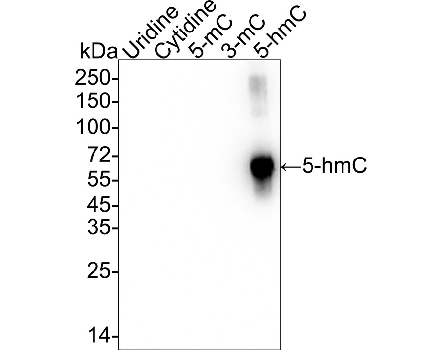
Western blot analysis of 5-Hydroxymethylcytosine (5-hmC) on different conjugations with Mouse anti-5-Hydroxymethylcytosine (5-hmC) antibody at 1/1,000 dilution. Lane 1: Uridine-BSA (negative), Lane 2: Cytidine-BSA (negative), Lane 3: 5-Methylcytosine-BSA (negative), Lane 4: 3-Methylcytosine-BSA (negative), Lane 5: 5-Hydroxymethylcytidine-BSA (positive), Lysates/proteins at 2 μg/Lane. Exposure time: 6 seconds; 4-20% SDS-PAGE gel. Proteins were transferred to a PVDF membrane and blocked with 5% NFDM/TBST for 1 hour at room temperature. The primary antibody at 1/1,000 dilution was used in 5% NFDM/TBST at room temperature for 2 hours. Goat Anti-Mouse IgG - HRP Secondary Antibody at 1/50,000 dilution was used for 1 hour at room temperature.IHC
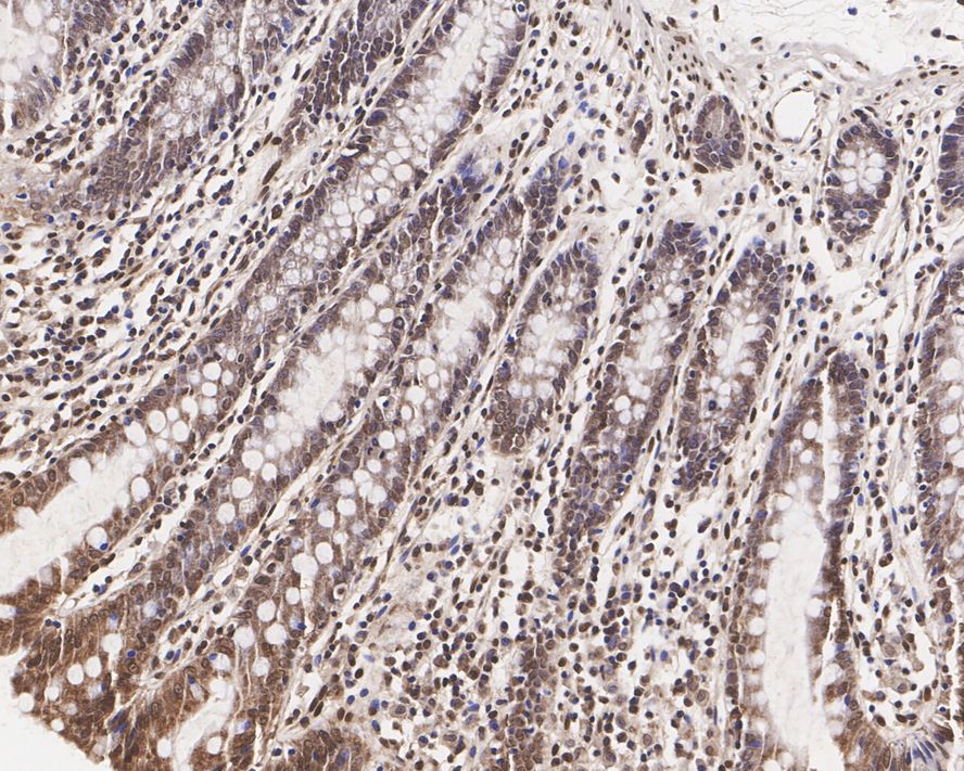
Immunohistochemical analysis of paraffin-embedded human colon tissue with Mouse anti-5-Hydroxymethylcytosine (5-hmC) antibody at 1/200 dilution. The section was denatured in 2M HCl for 15 minutes at room temperature, then neutralized in 0.1M Tris-HCl (pH 8.3) for 10 minutes. The tissues were blocked in 1% BSA for 20 minutes at room temperature, washed with ddH2O and PBS, and then probed with the primary antibody at 1/200 dilution for 1 hour at room temperature. The detection was performed using an HRP conjugated compact polymer system. DAB was used as the chromogen. Tissues were counterstained with hematoxylin and mounted with DPX.IHC
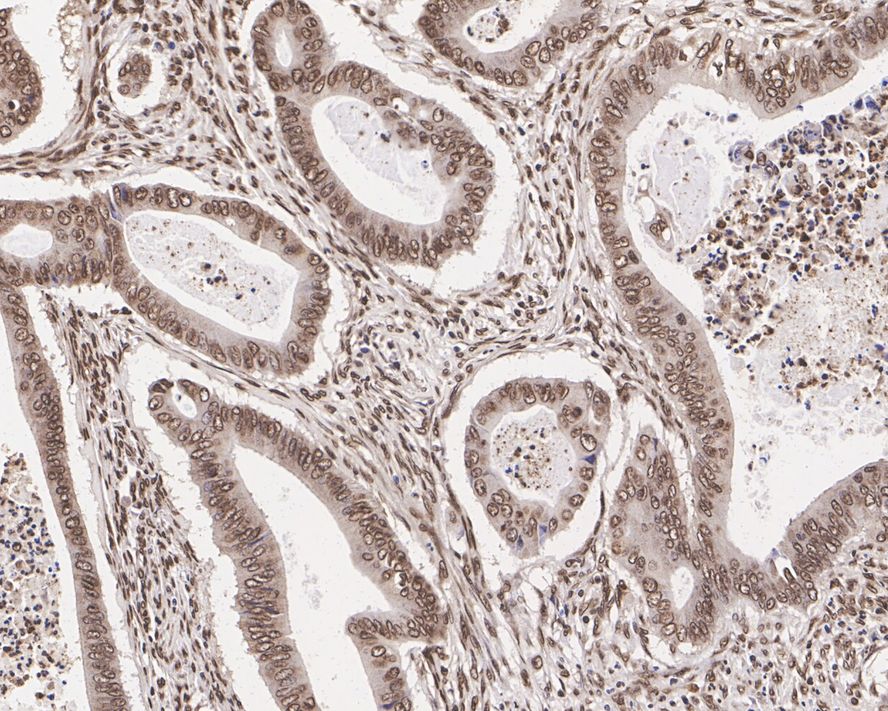
Immunohistochemical analysis of paraffin-embedded human colon cancer tissue with Mouse anti-5-Hydroxymethylcytosine (5-hmC) antibody at 1/200 dilution. The section was denatured in 2M HCl for 15 minutes at room temperature, then neutralized in 0.1M Tris-HCl (pH 8.3) for 10 minutes. The tissues were blocked in 1% BSA for 20 minutes at room temperature, washed with ddH2O and PBS, and then probed with the primary antibody at 1/200 dilution for 1 hour at room temperature. The detection was performed using an HRP conjugated compact polymer system. DAB was used as the chromogen. Tissues were counterstained with hematoxylin and mounted with DPX.IF
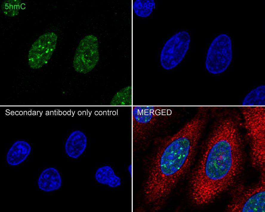
Immunocytochemistry analysis of HeLa cells labeling 5-Hydroxymethylcytosine (5-hmC) with Mouse anti-5-Hydroxymethylcytosine (5-hmC) antibody at 1/100 dilution. Cells were fixed in 70% ethyl alcohol for 5 minutes at room temperature, then subjected to acid hydrolysis using 2M HCl in TBST for 30 minutes at room temperature. Permeabilized with 0.1% Triton X-100 in PBS for 15 minutes, and then blocked with 2% BSA for 30 minutes at room temperature. Cells were then incubated with Mouse anti-5-Hydroxymethylcytosine (5-hmC) antibody at 1/100 dilution in 1% BSA in PBST overnight at 4 ℃. Goat Anti-Mouse IgG H&L (488) was used as the secondary antibody at 1/1,000 dilution. PBS instead of the primary antibody was used as the secondary antibody only control. Nuclear DNA was labelled in blue with DAPI. beta Tubulin (red) was stained at 1/100 dilution overnight at +4℃. Goat Anti-Rabbit IgG H&L (594) was used as the secondary antibody at 1/1,000 dilution.DB
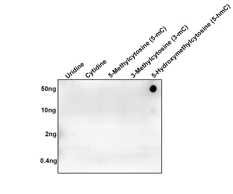
Dot blot analysis of 5-Hydroxymethylcytosine (5-hmC) on different conjugations with Mouse anti-5-Hydroxymethylcytosine (5-hmC) antibody at 1/1,000 dilution. Goat Anti-Mouse IgG - HRP Secondary Antibody at 1/50,000 dilution for 1 hour at room temperature. Lane 1: Uridine-BSA (negative), Lane 2: Cytidine-BSA (negative), Lane 3: 5-Methylcytosine-BSA (negative), Lane 4: 3-Methylcytosine-BSA (negative), Lane 5: 5-Hydroxymethylcytidine-BSA (positive), Proteins loading: 50ng, 10ng, 2ng, 0.4ng; Blocking and dilution buffer: 5% NFDM/TBST; Exposure time: 3 minutes.MeDIP
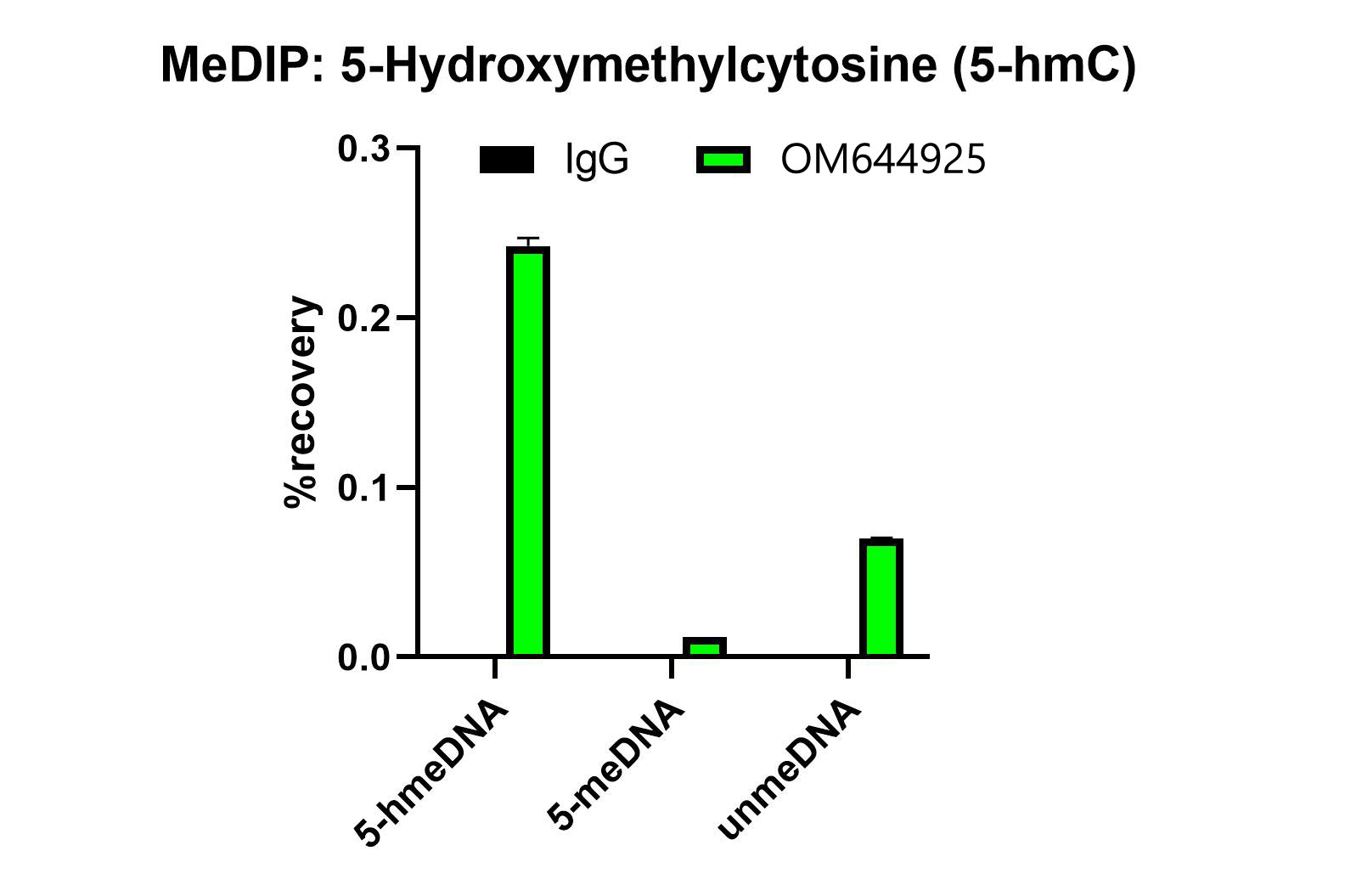
MeDIP was performed using anti-5-hmC antibody or Normal Mouse IgG. The same amount of unmethylated, 5-Methylcytosine (5-mC) or 5-Hydroxymethylcytosine (5-hmC) DNA standard was spiked in 1ug of genomic DNA isolated from HeLa cells as the control. Realtime PCR was then performed to determine the capture of DNA standard as in % of recovery, which is equivalent to one.| Product Name | 5-Hydroxymethylcytosine (5-hmC) Recombinant Mouse Monoclonal Antibody |
|---|---|
| Antibody Type | Primary Antibodies |
| Immunogen | Recombinant protein fused with 5-Hydroxymethylcytosine. |
| Clonality | monoclonal |
|---|---|
| Isotype | IgG1 |
| Host Species | Mouse |
| Tested Applications | DBICC/IFIHCMeDIPWB |
| WB:1:1000 IHC:1:200 ICC/IF:1:100 DB:1:1000 MeDIP:Use 5 μg for 3 μg of chromatin. |
|
| Species Reactivity | All |
| Concentration | 1mg/ml |
| Purification | Protein A |
| Alternative Names | 5-hmC antibody 5-hydroxymethylcytosine antibody 5hmC antibody |
|---|---|
| Molecular Weight(MW) | 66kDa |
| Function | 5-Hydroxymethylcytosine (5hmC) is a DNA pyrimidine nitrogen base derived from cytosine. It is potentially important in epigenetics, because the hydroxymethyl group on the cytosine can possibly switch a gene on and off. It was first seen in bacteriophages in 1952. However, in 2009 it was found to be abundant in human and mouse brains, as well as in embryonic stem cells. In mammals, it can be generated by oxidation of 5-methylcytosine, a reaction mediated by TET enzymes. |
WB
Western blot analysis of 5-Hydroxymethylcytosine (5-hmC) on different conjugations with Mouse anti-5-Hydroxymethylcytosine (5-hmC) antibody at 1/1,000 dilution. Lane 1: Uridine-BSA (negative), Lane 2: Cytidine-BSA (negative), Lane 3: 5-Methylcytosine-BSA (negative), Lane 4: 3-Methylcytosine-BSA (negative), Lane 5: 5-Hydroxymethylcytidine-BSA (positive), Lysates/proteins at 2 μg/Lane. Exposure time: 6 seconds; 4-20% SDS-PAGE gel. Proteins were transferred to a PVDF membrane and blocked with 5% NFDM/TBST for 1 hour at room temperature. The primary antibody at 1/1,000 dilution was used in 5% NFDM/TBST at room temperature for 2 hours. Goat Anti-Mouse IgG - HRP Secondary Antibody at 1/50,000 dilution was used for 1 hour at room temperature.
IHC
Immunohistochemical analysis of paraffin-embedded human colon tissue with Mouse anti-5-Hydroxymethylcytosine (5-hmC) antibody at 1/200 dilution. The section was denatured in 2M HCl for 15 minutes at room temperature, then neutralized in 0.1M Tris-HCl (pH 8.3) for 10 minutes. The tissues were blocked in 1% BSA for 20 minutes at room temperature, washed with ddH2O and PBS, and then probed with the primary antibody at 1/200 dilution for 1 hour at room temperature. The detection was performed using an HRP conjugated compact polymer system. DAB was used as the chromogen. Tissues were counterstained with hematoxylin and mounted with DPX.
IHC
Immunohistochemical analysis of paraffin-embedded human colon cancer tissue with Mouse anti-5-Hydroxymethylcytosine (5-hmC) antibody at 1/200 dilution. The section was denatured in 2M HCl for 15 minutes at room temperature, then neutralized in 0.1M Tris-HCl (pH 8.3) for 10 minutes. The tissues were blocked in 1% BSA for 20 minutes at room temperature, washed with ddH2O and PBS, and then probed with the primary antibody at 1/200 dilution for 1 hour at room temperature. The detection was performed using an HRP conjugated compact polymer system. DAB was used as the chromogen. Tissues were counterstained with hematoxylin and mounted with DPX.
IF
Immunocytochemistry analysis of HeLa cells labeling 5-Hydroxymethylcytosine (5-hmC) with Mouse anti-5-Hydroxymethylcytosine (5-hmC) antibody at 1/100 dilution. Cells were fixed in 70% ethyl alcohol for 5 minutes at room temperature, then subjected to acid hydrolysis using 2M HCl in TBST for 30 minutes at room temperature. Permeabilized with 0.1% Triton X-100 in PBS for 15 minutes, and then blocked with 2% BSA for 30 minutes at room temperature. Cells were then incubated with Mouse anti-5-Hydroxymethylcytosine (5-hmC) antibody at 1/100 dilution in 1% BSA in PBST overnight at 4 ℃. Goat Anti-Mouse IgG H&L (488) was used as the secondary antibody at 1/1,000 dilution. PBS instead of the primary antibody was used as the secondary antibody only control. Nuclear DNA was labelled in blue with DAPI. beta Tubulin (red) was stained at 1/100 dilution overnight at +4℃. Goat Anti-Rabbit IgG H&L (594) was used as the secondary antibody at 1/1,000 dilution.
DB
Dot blot analysis of 5-Hydroxymethylcytosine (5-hmC) on different conjugations with Mouse anti-5-Hydroxymethylcytosine (5-hmC) antibody at 1/1,000 dilution. Goat Anti-Mouse IgG - HRP Secondary Antibody at 1/50,000 dilution for 1 hour at room temperature. Lane 1: Uridine-BSA (negative), Lane 2: Cytidine-BSA (negative), Lane 3: 5-Methylcytosine-BSA (negative), Lane 4: 3-Methylcytosine-BSA (negative), Lane 5: 5-Hydroxymethylcytidine-BSA (positive), Proteins loading: 50ng, 10ng, 2ng, 0.4ng; Blocking and dilution buffer: 5% NFDM/TBST; Exposure time: 3 minutes.
MeDIP
MeDIP was performed using anti-5-hmC antibody or Normal Mouse IgG. The same amount of unmethylated, 5-Methylcytosine (5-mC) or 5-Hydroxymethylcytosine (5-hmC) DNA standard was spiked in 1ug of genomic DNA isolated from HeLa cells as the control. Realtime PCR was then performed to determine the capture of DNA standard as in % of recovery, which is equivalent to one.| Application Notes | WB:1:1000 IHC:1:200 ICC/IF:1:100 DB:1:1000 MeDIP:Use 5 μg for 3 μg of chromatin. |
|---|
| Form | Liquid |
|---|---|
| Storage Instructions | Store at +4℃ after thawing. Aliquot store at -20℃. Avoid repeated freeze / thaw cycles. |
| Storage Buffer | 1*TBS (pH7.4), 0.05% BSA, 40% Glycerol. Preservative: 0.05% Sodium Azide. |


