
WB
Western blot analysis of anti-TRAC antibody. The sample well of each lane was loaded with 30 ug of sample under reducing conditions. Lane 1: human Jurkat whole cell lysates. After electrophoresis, proteins were transferred to a membrane. Then the membrane was incubated with rabbit anti-TRAC antigen affinity purified polyclonal antibody and probed with a goat anti-rabbit IgG-HRP secondary antibody. The signal is developed using ECL Plus Western Blotting Substrate.IHC
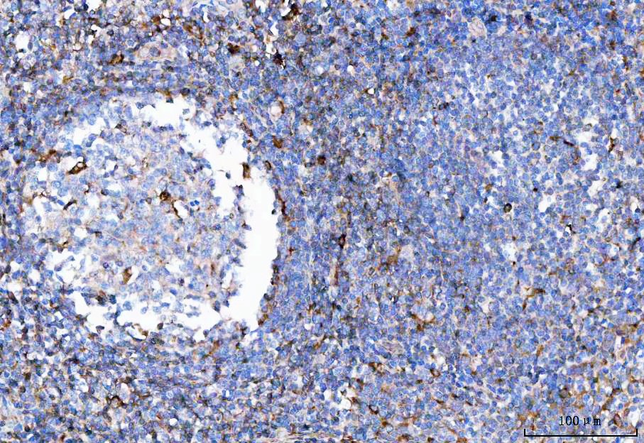
IHC analysis of TRAC using anti-TRAC antibody. TRAC was detected in a paraffin-embedded section of human tonsil tissue. The tissue section was developed using HRP Conjugated Rabbit IgG Super Vision Assay Kit with DAB as the chromogen.IHC
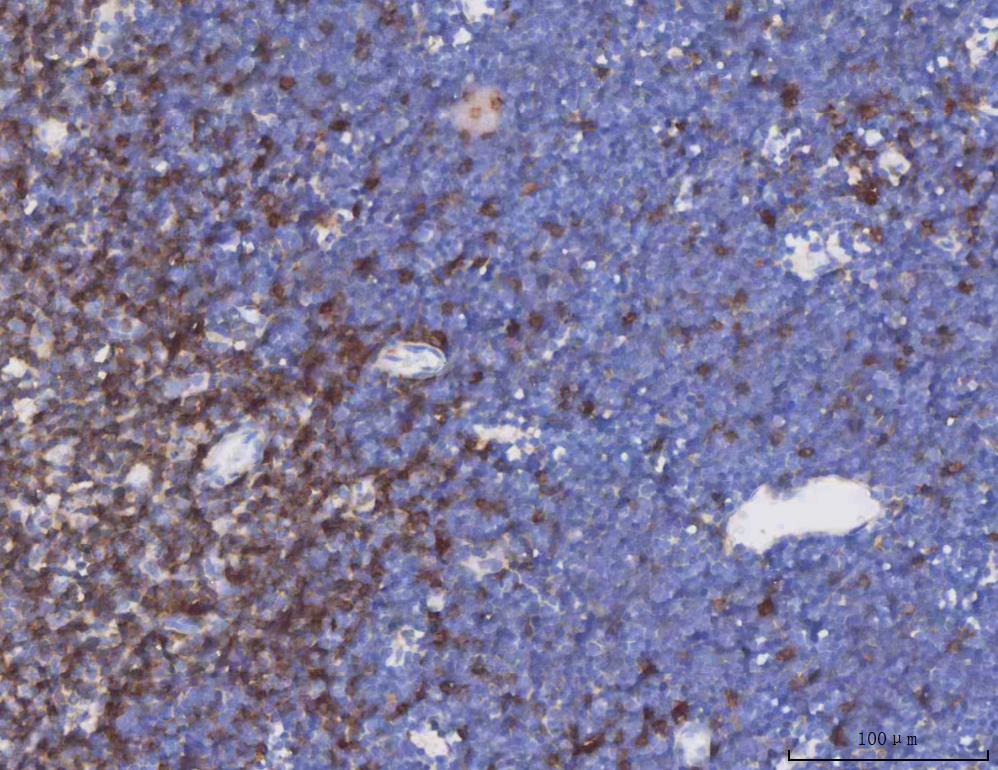
IHC analysis of TRAC using anti-TRAC antibody. TRAC was detected in a paraffin-embedded section of mouse thymus tissue. The tissue section was developed using HRP Conjugated Rabbit IgG Super Vision Assay Kit with DAB as the chromogen.IHC
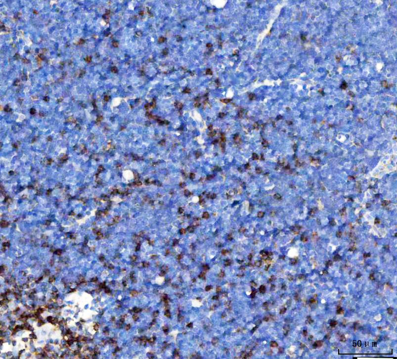
IHC analysis of TRAC using anti-TRAC antibody. TRAC was detected in a paraffin-embedded section of mouse thymus tissue. The tissue section was developed using HRP Conjugated Rabbit IgG Super Vision Assay Kit with DAB as the chromogen.IHC
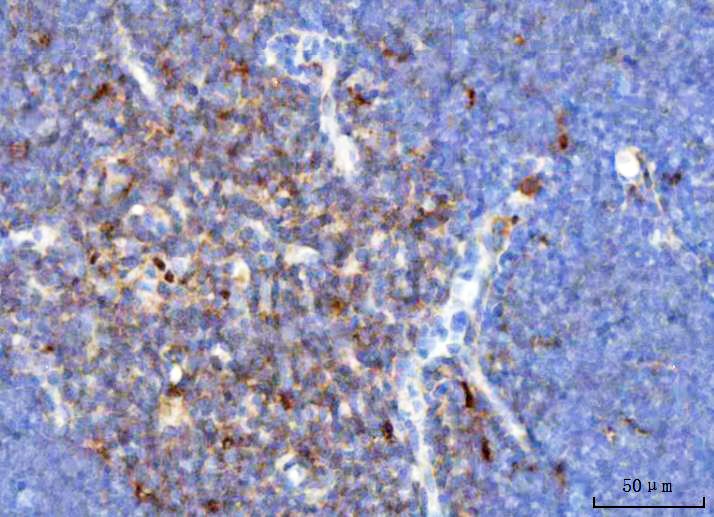
IHC analysis of TRAC using anti-TRAC antibody. TRAC was detected in a paraffin-embedded section of rat thymus tissue. The tissue section was developed using HRP Conjugated Rabbit IgG Super Vision Assay Kitwith DAB as the chromogen.FC
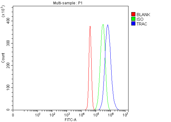
Flow Cytometry analysis of Jurkat cells using anti-TRAC antibody. Overlay histogram showing Jurkat cells stained with anti-TRAC antibody (Blue line). The cells were fixed with 4% paraformaldehyde and blocked with 10% normal goat serum. And then incubated with rabbit anti-TRAC Antibody (1:100). DyLight®488 conjugated goat anti-rabbit IgG (1:100) was used as secondary antibody. Isotype control antibody (Green line) was rabbit IgG (1:100) used under the same conditions. Unlabelled sample (Red line) was also used as a control.| Product Name | Rabbit polyclonal antibody to TRAC |
|---|---|
| Antibody Type | Primary Antibodies |
| Immunogen | E. coli-derived human TCR alpha recombinant protein (Position: I1-L114). Human TCR alpha shares 57.8% amino acid (aa) sequence identity with mouse TCR alpha. |
| Clonality | Polyclonal |
|---|---|
| Isotype | IgG |
| Host Species | Rabbit |
| Tested Applications | FCIHCWB |
| WB:1:500-1:2000 IHC:1:50-1:400 FC:1:50-1:200 |
|
| Species Reactivity | HumanMouseRat |
| Concentration | 0.5mg/ml |
| Purification | Affinity purified |
| Gene Symbol | TRAC |
|---|---|
| Gene Synonyms | TRA IMD7 TCRA TRCA |
| Gene Full Name | T cell receptor alpha constant |
| Gene Summary | T cell receptors recognize foreign antigens which have been processed as small peptides and bound to major histocompatibility complex (MHC) molecules at the surface of antigen presenting cells (APC). Each T cell receptor is a dimer consisting of one alpha and one beta chain or one delta and one gamma chain. In a single cell, the T cell receptor loci are rearranged and expressed in the order delta, gamma, beta, and alpha. If both delta and gamma rearrangements produce functional chains, the cell expresses delta and gamma. If not, the cell proceeds to rearrange the beta and alpha loci. This region represents the germline organization of the T cell receptor alpha and delta loci. Both the alpha and delta loci include V (variable), J (joining), and C (constant) segments and the delta locus also includes diversity (D) segments. The delta locus is situated within the alpha locus, between the alpha V and J segments. During T cell development, the delta chain is synthesized by a recombination event at the DNA level joining a D segment with a J segment; a V segment is then joined to the D-J gene. The alpha chain is synthesized by recombination joining a single V segment with a J segment. For both chains, the C segment is later joined by splicing at the RNA level. Recombination of many different V segments with several J segments provides a wide range of antigen recognition. Additional diversity is attained by junctional diversity, resulting from the random additional of nucleotides by terminal deoxynucleotidyltransferase. Five variable segments can be used in either alpha or delta chains and are described by TRAV/DV symbols. Several V and J segments of the alpha locus are known to be incapable of encoding a protein and are considered pseudogenes. [provided by RefSeq, Aug 2016] |
| Alternative Names | T cell receptor alpha constant IMD7 TCRA TRA TRCA TRAC |
| Molecular Weight(MW) | 45 kDa |
| Cellular Localization | Cell membrane. |
WB
Western blot analysis of anti-TRAC antibody. The sample well of each lane was loaded with 30 ug of sample under reducing conditions. Lane 1: human Jurkat whole cell lysates. After electrophoresis, proteins were transferred to a membrane. Then the membrane was incubated with rabbit anti-TRAC antigen affinity purified polyclonal antibody and probed with a goat anti-rabbit IgG-HRP secondary antibody. The signal is developed using ECL Plus Western Blotting Substrate.
IHC
IHC analysis of TRAC using anti-TRAC antibody. TRAC was detected in a paraffin-embedded section of human tonsil tissue. The tissue section was developed using HRP Conjugated Rabbit IgG Super Vision Assay Kit with DAB as the chromogen.
IHC
IHC analysis of TRAC using anti-TRAC antibody. TRAC was detected in a paraffin-embedded section of mouse thymus tissue. The tissue section was developed using HRP Conjugated Rabbit IgG Super Vision Assay Kit with DAB as the chromogen.
IHC
IHC analysis of TRAC using anti-TRAC antibody. TRAC was detected in a paraffin-embedded section of mouse thymus tissue. The tissue section was developed using HRP Conjugated Rabbit IgG Super Vision Assay Kit with DAB as the chromogen.
IHC
IHC analysis of TRAC using anti-TRAC antibody. TRAC was detected in a paraffin-embedded section of rat thymus tissue. The tissue section was developed using HRP Conjugated Rabbit IgG Super Vision Assay Kitwith DAB as the chromogen.
FC
Flow Cytometry analysis of Jurkat cells using anti-TRAC antibody. Overlay histogram showing Jurkat cells stained with anti-TRAC antibody (Blue line). The cells were fixed with 4% paraformaldehyde and blocked with 10% normal goat serum. And then incubated with rabbit anti-TRAC Antibody (1:100). DyLight®488 conjugated goat anti-rabbit IgG (1:100) was used as secondary antibody. Isotype control antibody (Green line) was rabbit IgG (1:100) used under the same conditions. Unlabelled sample (Red line) was also used as a control.| Application Notes | WB:1:500-1:2000 IHC:1:50-1:400 FC:1:50-1:200 |
|---|
| Form | Liquid |
|---|---|
| Storage Instructions | 12 months from date of receipt, -20℃ as supplied. 6 months 2 to 8℃ after reconstitution. Avoid repeated freezing and thawing. |
| Storage Buffer | 500ug/ml antibody with PBS, 0.02% NaN3, 1 mg/ml BSA and 50% glycerol. |


