WB
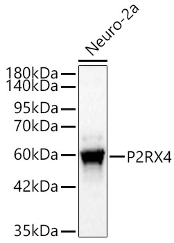
Western blot analysis of Neuro-2a cells using P2RX4 Rabbit mAb at 1:1000 dilution. Secondary antibody: HRP-conjugated Goat anti-Rabbit IgG (H+L) at 1:10000 dilution. Lysates/proteins: 25 μg per lane. Blocking buffer: 3% nonfat dry milk in TBST. Detection: ECL Basic Kit. Exposure time: 30s.IHC
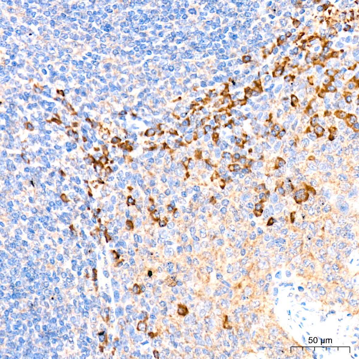
Immunohistochemistry analysis of paraffin embedded Human tonsil tissue using P2RX4 Rabbit mAb at a dilution of 1:200 (40x lens). High pressure antigen retrieval was performed with 0.01 M citrate buffer (pH 6.0) prior to IHC staining.IHC
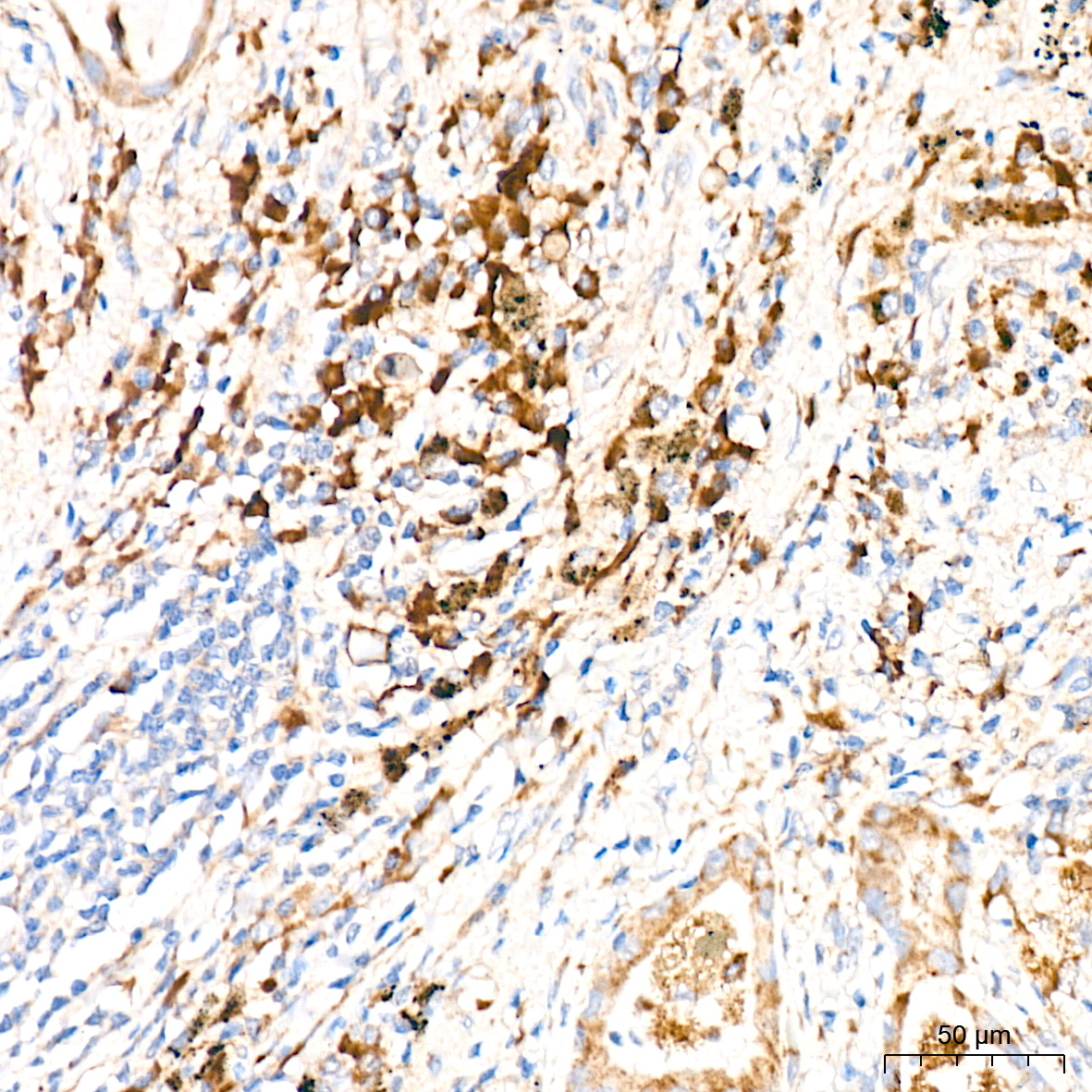
Immunohistochemistry analysis of paraffin embedded Human lung tissue using P2RX4 Rabbit mAb at a dilution of 1:200 (40x lens). High pressure antigen retrieval was performed with 0.01 M citrate buffer (pH 6.0) prior to IHC staining.ICC/IF
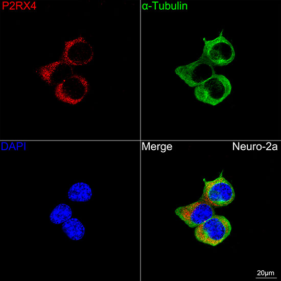
Confocal imaging of Neuro-2a cells using P2RX4 Rabbit mAb ( dilution 1:200) followed by a further incubation with Cy3 Goat Anti-Rabbit IgG (H+L) (dilution 1:500) (Red). The cells were counterstained with α-Tubulin Mouse mAb (dilution 1:400) followed by incubation withOmnimabs® 488 Goat Anti-Mouse IgG(H&L) (dilution 1:500) (Green). DAPI was used for nuclear staining (Blue). Objective: 100x.IF-P
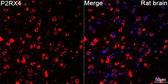
Confocal imaging of paraffin-embedded Rat brain tissue using P2RX4 Rabbit mAb(dilution 1:200) followed by a further incubation with Cy3 Goat Anti-Rabbit IgG (H+L) (dilution 1:500) (Red). DAPI was used for nuclear staining (Blue). Objective: 40x. Perform microwave antigen retrieval with 0.01 M citrate buffer (pH 6.0) prior to IF staining.| Product Name | P2RX4 Rabbit mAb |
|---|---|
| Antibody Type | Primary Antibodies |
| Immunogen | Recombinant protein. |
| Clonality | Monoclonal |
|---|---|
| Isotype | IgG |
| Host Species | Rabbit |
| Tested Applications | ICC/IFIF-PIHCWB |
| WB:1:500-1:1000 IHC:1:50-1:200 ICC/IF:1:50-1:200 IF-P:1:50-1:200 |
|
| Species Reactivity | HumanMouseRat |
| Concentration | 1mg/ml |
| Purification | Affinity purified |
| Gene Symbol | P2RX4 |
|---|---|
| Gene Synonyms | P2X4 P2X4R |
| Gene Full Name | purinergic receptor P2X 4 |
| Gene Summary | The product of this gene belongs to the family of purinoceptors for ATP. This receptor functions as a ligand-gated ion channel with high calcium permeability. The main pharmacological distinction between the members of the purinoceptor family is the relative sensitivity to the antagonists suramin and PPADS. The product of this gene has the lowest sensitivity for these antagonists. Multiple alternatively spliced transcript variants, some protein-coding and some not protein-coding, have been found for this gene. [provided by RefSeq, Feb 2012] |
| Alternative Names | P2X4 P2X4R P2RX4 |
| Molecular Weight(MW) | 43kDa(Observed MW 43-62kDa) |
| Cellular Localization | Membrane, Multi-pass membrane protein. |
WB
Western blot analysis of Neuro-2a cells using P2RX4 Rabbit mAb at 1:1000 dilution. Secondary antibody: HRP-conjugated Goat anti-Rabbit IgG (H+L) at 1:10000 dilution. Lysates/proteins: 25 μg per lane. Blocking buffer: 3% nonfat dry milk in TBST. Detection: ECL Basic Kit. Exposure time: 30s.
IHC
Immunohistochemistry analysis of paraffin embedded Human tonsil tissue using P2RX4 Rabbit mAb at a dilution of 1:200 (40x lens). High pressure antigen retrieval was performed with 0.01 M citrate buffer (pH 6.0) prior to IHC staining.
IHC
Immunohistochemistry analysis of paraffin embedded Human lung tissue using P2RX4 Rabbit mAb at a dilution of 1:200 (40x lens). High pressure antigen retrieval was performed with 0.01 M citrate buffer (pH 6.0) prior to IHC staining.
ICC/IF
Confocal imaging of Neuro-2a cells using P2RX4 Rabbit mAb ( dilution 1:200) followed by a further incubation with Cy3 Goat Anti-Rabbit IgG (H+L) (dilution 1:500) (Red). The cells were counterstained with α-Tubulin Mouse mAb (dilution 1:400) followed by incubation withOmnimabs® 488 Goat Anti-Mouse IgG(H&L) (dilution 1:500) (Green). DAPI was used for nuclear staining (Blue). Objective: 100x.
IF-P
Confocal imaging of paraffin-embedded Rat brain tissue using P2RX4 Rabbit mAb(dilution 1:200) followed by a further incubation with Cy3 Goat Anti-Rabbit IgG (H+L) (dilution 1:500) (Red). DAPI was used for nuclear staining (Blue). Objective: 40x. Perform microwave antigen retrieval with 0.01 M citrate buffer (pH 6.0) prior to IF staining.| Application Notes | WB:1:500-1:1000 IHC:1:50-1:200 ICC/IF:1:50-1:200 IF-P:1:50-1:200 |
|---|
| Form | Liquid |
|---|---|
| Storage Instructions | Store at -20℃. Avoid freeze / thaw cycles. |
| Storage Buffer | PBS containing 50% glycerol and 0.05% BSA, preserved with proclin300 or sodium azide (as specified on the Certificate of Analysis), pH 7.3. |


