WB
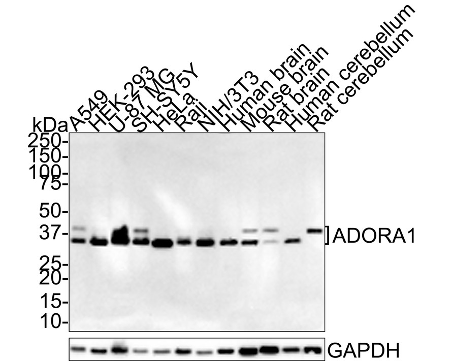
Western blot analysis of Adenosine A1 Receptor on different lysates with Rabbit anti-Adenosine A1 Receptor antibody at 1/1,000 dilution. Lane 1: A549 cell lysate (15 µg/Lane), Lane 2: HEK-293 cell lysate (15 µg/Lane), Lane 3: U-87 MG cell lysate (15 µg/Lane), Lane 4: SH-SY5Y cell lysate (15 µg/Lane), Lane 5: HeLa cell lysate (15 µg/Lane), Lane 6: Raji cell lysate (15 µg/Lane), Lane 7: NIH/3T3 cell lysate (15 µg/Lane), Lane 8: Human brain tissue lysate (30 µg/Lane), Lane 9: Mouse brain tissue lysate (30 µg/Lane), Lane 10: Rat brain tissue lysate (30 µg/Lane), Lane 11: Human cerebellum tissue lysate (30 µg/Lane), Lane 12: Rat cerebellum tissue lysate (30 µg/Lane), Exposure time: 5 minutes; 4-20% SDS-PAGE gel. Proteins were transferred to a PVDF membrane and blocked with 5% NFDM/TBST for 1 hour at room temperature. The primary antibody at 1/1,000 dilution was used in 5% NFDM/TBST at room temperature for 2 hours. Goat Anti-Rabbit IgG - HRP Secondary Antibody at 1:100,000 dilution was used for 1 hour at room temperature.IHC
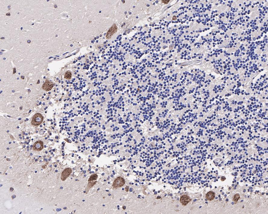
Immunohistochemical analysis of paraffin-embedded human cerebellum tissue with Rabbit anti-Adenosine A1 Receptor antibody at 1/1,000 dilution. The section was pre-treated using heat mediated antigen retrieval with Tris-EDTA buffer (pH 9.0) for 20 minutes. The tissues were blocked in 1% BSA for 20 minutes at room temperature, washed with ddH2O and PBS, and then probed with the primary antibody at 1/1,000 dilution for 1 hour at room temperature. The detection was performed using an HRP conjugated compact polymer system. DAB was used as the chromogen. Tissues were counterstained with hematoxylin and mounted with DPX.IHC
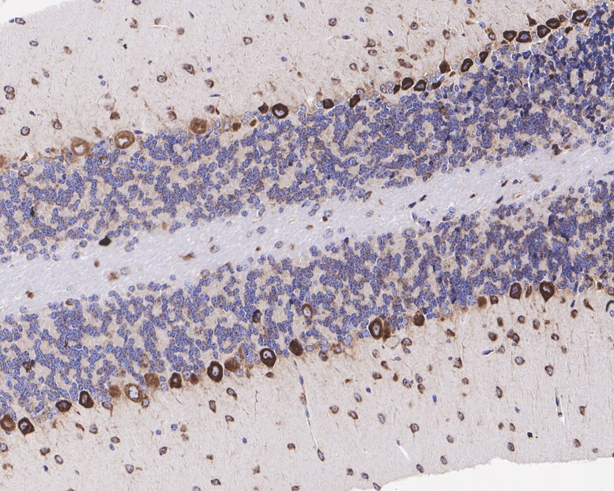
Immunohistochemical analysis of paraffin-embedded mouse cerebellum tissue with Rabbit anti-Adenosine A1 Receptor antibody at 1/1,000 dilution. The section was pre-treated using heat mediated antigen retrieval with Tris-EDTA buffer (pH 9.0) for 20 minutes. The tissues were blocked in 1% BSA for 20 minutes at room temperature, washed with ddH2O and PBS, and then probed with the primary antibody at 1/1,000 dilution for 1 hour at room temperature. The detection was performed using an HRP conjugated compact polymer system. DAB was used as the chromogen. Tissues were counterstained with hematoxylin and mounted with DPX.IHC
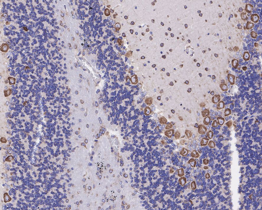
Immunohistochemical analysis of paraffin-embedded rat cerebellum tissue with Rabbit anti-Adenosine A1 Receptor antibody at 1/1,000 dilution. The section was pre-treated using heat mediated antigen retrieval with Tris-EDTA buffer (pH 9.0) for 20 minutes. The tissues were blocked in 1% BSA for 20 minutes at room temperature, washed with ddH2O and PBS, and then probed with the primary antibody at 1/1,000 dilution for 1 hour at room temperature. The detection was performed using an HRP conjugated compact polymer system. DAB was used as the chromogen. Tissues were counterstained with hematoxylin and mounted with DPX.| Product Name | Adenosine A1 Receptor Recombinant Rabbit Monoclonal Antibody |
|---|---|
| Antibody Type | Primary Antibodies |
| Immunogen | Recombinant protein within Human Adenosine A1 Receptor aa 103-201 / 326. |
| Clonality | monoclonal |
|---|---|
| Isotype | IgG |
| Host Species | Rabbit |
| Tested Applications | IHCWB |
| WB:1:1000 IHC:1:1000 |
|
| Species Reactivity | HumanMouseRat |
| Concentration | 1mg/ml |
| Purification | Protein A |
| Gene Symbol | ADORA1 |
|---|---|
| Gene Synonyms | RDC7 |
| Gene Full Name | adenosine A1 receptor |
| Gene Summary | The protein encoded by this gene is an adenosine receptor that belongs to the G-protein coupled receptor 1 family. There are 3 types of adenosine receptors, each with a specific pattern of ligand binding and tissue distribution, and together they regulate a diverse set of physiologic functions. The type A1 receptors inhibit adenylyl cyclase, and play a role in the fertilization process. Animal studies also suggest a role for A1 receptors in kidney function and ethanol intoxication. Transcript variants with alternative splicing in the 5' UTR have been found for this gene. [provided by RefSeq, Jul 2008] |
| Alternative Names | A1AR antibody A1R antibody AA1R antibody AA1R_HUMAN antibody Adenosine receptor A1 antibody ADORA 1 antibody ADORA 1 antibody ADORA1 antibody RDC 7 antibody RDC7 antibody A1AR antibody A1R antibody AA1R antibody AA1R_HUMAN antibody Adenosine receptor A1 antibody ADORA 1 antibody ADORA 1 antibody ADORA1 antibody RDC 7 antibody RDC7 antibody Ri antibody |
| Molecular Weight(MW) | 36.5/40kDa |
| Cellular Localization | Cell membrane. |
WB
Western blot analysis of Adenosine A1 Receptor on different lysates with Rabbit anti-Adenosine A1 Receptor antibody at 1/1,000 dilution. Lane 1: A549 cell lysate (15 µg/Lane), Lane 2: HEK-293 cell lysate (15 µg/Lane), Lane 3: U-87 MG cell lysate (15 µg/Lane), Lane 4: SH-SY5Y cell lysate (15 µg/Lane), Lane 5: HeLa cell lysate (15 µg/Lane), Lane 6: Raji cell lysate (15 µg/Lane), Lane 7: NIH/3T3 cell lysate (15 µg/Lane), Lane 8: Human brain tissue lysate (30 µg/Lane), Lane 9: Mouse brain tissue lysate (30 µg/Lane), Lane 10: Rat brain tissue lysate (30 µg/Lane), Lane 11: Human cerebellum tissue lysate (30 µg/Lane), Lane 12: Rat cerebellum tissue lysate (30 µg/Lane), Exposure time: 5 minutes; 4-20% SDS-PAGE gel. Proteins were transferred to a PVDF membrane and blocked with 5% NFDM/TBST for 1 hour at room temperature. The primary antibody at 1/1,000 dilution was used in 5% NFDM/TBST at room temperature for 2 hours. Goat Anti-Rabbit IgG - HRP Secondary Antibody at 1:100,000 dilution was used for 1 hour at room temperature.
IHC
Immunohistochemical analysis of paraffin-embedded human cerebellum tissue with Rabbit anti-Adenosine A1 Receptor antibody at 1/1,000 dilution. The section was pre-treated using heat mediated antigen retrieval with Tris-EDTA buffer (pH 9.0) for 20 minutes. The tissues were blocked in 1% BSA for 20 minutes at room temperature, washed with ddH2O and PBS, and then probed with the primary antibody at 1/1,000 dilution for 1 hour at room temperature. The detection was performed using an HRP conjugated compact polymer system. DAB was used as the chromogen. Tissues were counterstained with hematoxylin and mounted with DPX.
IHC
Immunohistochemical analysis of paraffin-embedded mouse cerebellum tissue with Rabbit anti-Adenosine A1 Receptor antibody at 1/1,000 dilution. The section was pre-treated using heat mediated antigen retrieval with Tris-EDTA buffer (pH 9.0) for 20 minutes. The tissues were blocked in 1% BSA for 20 minutes at room temperature, washed with ddH2O and PBS, and then probed with the primary antibody at 1/1,000 dilution for 1 hour at room temperature. The detection was performed using an HRP conjugated compact polymer system. DAB was used as the chromogen. Tissues were counterstained with hematoxylin and mounted with DPX.
IHC
Immunohistochemical analysis of paraffin-embedded rat cerebellum tissue with Rabbit anti-Adenosine A1 Receptor antibody at 1/1,000 dilution. The section was pre-treated using heat mediated antigen retrieval with Tris-EDTA buffer (pH 9.0) for 20 minutes. The tissues were blocked in 1% BSA for 20 minutes at room temperature, washed with ddH2O and PBS, and then probed with the primary antibody at 1/1,000 dilution for 1 hour at room temperature. The detection was performed using an HRP conjugated compact polymer system. DAB was used as the chromogen. Tissues were counterstained with hematoxylin and mounted with DPX.| Application Notes | WB:1:1000 IHC:1:1000 |
|---|
| Form | Liquid |
|---|---|
| Storage Instructions | Store at +4℃ after thawing. Aliquot store at -20℃. Avoid repeated freeze / thaw cycles. |
| Storage Buffer | 1*TBS (pH7.4), 0.05% BSA, 40% Glycerol. Preservative: 0.05% Sodium Azide. |


