WB
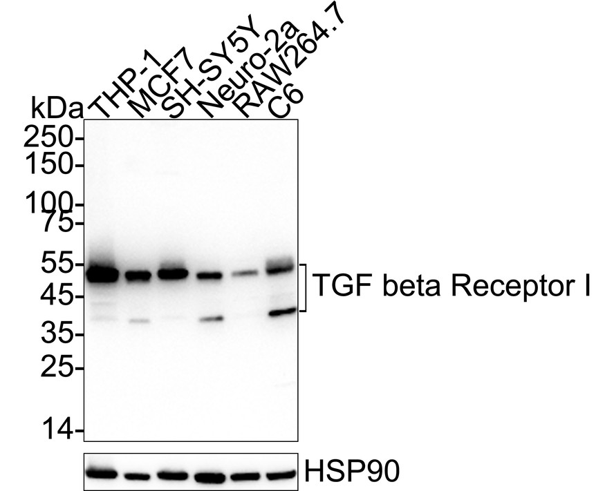
Western blot analysis of TGF beta Receptor I on different lysates with Rabbit anti-TGF beta Receptor I antibody at 1/2,000 dilution. Lane 1: THP-1 cell lysate, Lane 2: MCF7 cell lysate, Lane 3: SH-SY5Y cell lysate, Lane 4: Neuro-2a cell lysate, Lane 5: RAW264.7 cell lysate, Lane 6: C6 cell lysate, Lysates/proteins at 20 µg/Lane. Exposure time: 2 minutes; 4-20% SDS-PAGE gel. Proteins were transferred to a PVDF membrane and blocked with 5% NFDM/TBST for 1 hour at room temperature. The primary antibody at 1/2,000 dilution was used in primary antibody dilution at 4℃ overnight. Goat Anti-Rabbit IgG - HRP Secondary Antibody at 1/50,000 dilution was used for 1 hour at room temperature.IP
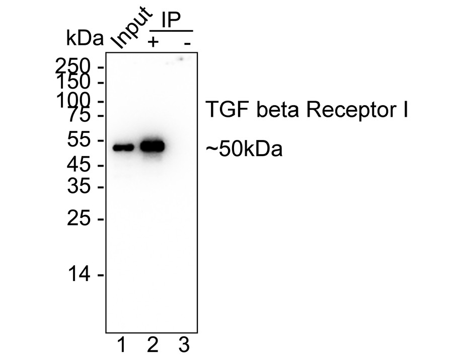
TGF beta Receptor I was immunoprecipitated from 0.2 mg THP-1 cell lysate with Rabbit anti-TGF beta Receptor I antibody at 2 µg/10 µl beads. Western blot was performed from the immunoprecipitate using Rabbit anti-TGF beta Receptor I antibody at 1/2,000 dilution. HRP Conjugated Anti-Rabbit IgG for IP Nano-secondary antibody at 1/5,000 dilution was used for 1 hour at room temperature. Lane 1: THP-1 cell lysate (input), Lane 2: Rabbit anti-TGF beta Receptor I antibody IP in THP-1 cell lysate, Lane 3: Rabbit IgG instead of Rabbit anti-TGF beta Receptor I antibody in THP-1 cell lysate, Blocking/Dilution buffer: 5% NFDM/TBST, Exposure time: 1 minute 7 seconds.| Product Name | TGF beta Receptor I Recombinant Rabbit Monoclonal Antibody |
|---|---|
| Antibody Type | Primary Antibodies |
| Immunogen | Recombinant protein |
| Clonality | monoclonal |
|---|---|
| Isotype | IgG |
| Host Species | Rabbit |
| Tested Applications | IPWB |
| WB:1:2000 IP:1-2μg/sample |
|
| Species Reactivity | HumanMouseRat |
| Concentration | 1mg/ml |
| Purification | Protein A |
| Gene Symbol | TGFBR1 |
|---|---|
| Gene Synonyms | AAT5 ALK5 ESS1 LDS1 MSSE SKR4 TBRI ALK-5 LDS1A LDS2A TBR-i TGFR-1 ACVRLK4 tbetaR-I |
| Gene Full Name | transforming growth factor beta receptor 1 |
| Gene Summary | The protein encoded by this gene forms a heteromeric complex with type II TGF-beta receptors when bound to TGF-beta, transducing the TGF-beta signal from the cell surface to the cytoplasm. The encoded protein is a serine/threonine protein kinase. Mutations in this gene have been associated with Loeys-Dietz aortic aneurysm syndrome (LDAS). Multiple transcript variants encoding different isoforms have been found for this gene. [provided by RefSeq, Aug 2008] |
| Alternative Names | AAT 5 antibody AAT5 antibody Activin A receptor type II like kinase 53kDa antibody Activin A receptor type II like kinase antibody Activin A receptor type II like kinase 53kD antibody Activin A receptor type II like protein kinase of 53kD antibody activin A receptor type II-like kinase 53kDa antibody activin A receptor type II-like protein kinase of 53kD antibody Activin receptor like kinase 5 antibody Activin receptor-like kinase 5 antibody AAT 5 antibody AAT5 antibody Activin A receptor type II like kinase 53kDa antibody Activin A receptor type II like kinase antibody Activin A receptor type II like kinase 53kD antibody Activin A receptor type II like protein kinase of 53kD antibody activin A receptor type II-like kinase 53kDa antibody activin A receptor type II-like protein kinase of 53kD antibody Activin receptor like kinase 5 antibody Activin receptor-like kinase 5 antibody ACVRLK 4 antibody ACVRLK4 antibody ALK 5 antibody ALK-5 antibody ALK5 antibody LDS1A antibody LDS2A antibody MSSE antibody Serine/threonine protein kinase receptor R4 antibody Serine/threonine-protein kinase receptor R4 antibody SKR 4 antibody SKR4 antibody TbetaR I antibody TbetaR-I antibody TGF beta receptor type 1 antibody TGF beta receptor type I antibody TGF beta type I receptor antibody TGF-beta receptor type I antibody TGF-beta receptor type-1 antibody TGF-beta type I receptor antibody TGFBR 1 antibody TGFBR1 antibody TGFBR1 protein antibody TGFR 1 antibody TGFR-1 antibody TGFR1 antibody TGFR1_HUMAN antibody Transforming growth factor beta receptor 1 antibody Transforming growth factor beta receptor I (activin A receptor type II like kinase 53kD) antibody Transforming growth factor beta receptor I antibody transforming growth factor beta receptor 1 antibody transforming growth factor beta receptor I (activin A receptor type II-like kinase 53kD) antibody Transforming growth factor-beta receptor type I antibody |
| Molecular Weight(MW) | 56kDa(Observed band size: 50/37kDa) |
| Cellular Localization | Cell membrane, Cell junction, tight junction, Cell surface, Membrane raft. |
WB
Western blot analysis of TGF beta Receptor I on different lysates with Rabbit anti-TGF beta Receptor I antibody at 1/2,000 dilution. Lane 1: THP-1 cell lysate, Lane 2: MCF7 cell lysate, Lane 3: SH-SY5Y cell lysate, Lane 4: Neuro-2a cell lysate, Lane 5: RAW264.7 cell lysate, Lane 6: C6 cell lysate, Lysates/proteins at 20 µg/Lane. Exposure time: 2 minutes; 4-20% SDS-PAGE gel. Proteins were transferred to a PVDF membrane and blocked with 5% NFDM/TBST for 1 hour at room temperature. The primary antibody at 1/2,000 dilution was used in primary antibody dilution at 4℃ overnight. Goat Anti-Rabbit IgG - HRP Secondary Antibody at 1/50,000 dilution was used for 1 hour at room temperature.
IP
TGF beta Receptor I was immunoprecipitated from 0.2 mg THP-1 cell lysate with Rabbit anti-TGF beta Receptor I antibody at 2 µg/10 µl beads. Western blot was performed from the immunoprecipitate using Rabbit anti-TGF beta Receptor I antibody at 1/2,000 dilution. HRP Conjugated Anti-Rabbit IgG for IP Nano-secondary antibody at 1/5,000 dilution was used for 1 hour at room temperature. Lane 1: THP-1 cell lysate (input), Lane 2: Rabbit anti-TGF beta Receptor I antibody IP in THP-1 cell lysate, Lane 3: Rabbit IgG instead of Rabbit anti-TGF beta Receptor I antibody in THP-1 cell lysate, Blocking/Dilution buffer: 5% NFDM/TBST, Exposure time: 1 minute 7 seconds.| Application Notes | WB:1:2000 IP:1-2μg/sample |
|---|
| Form | Liquid |
|---|---|
| Storage Instructions | Store at +4℃ after thawing. Aliquot store at -20℃. Avoid repeated freeze / thaw cycles. |
| Storage Buffer | PBS (pH7.4), 0.1% BSA, 40% Glycerol. Preservative: 0.05% Sodium Azide. |


