WB
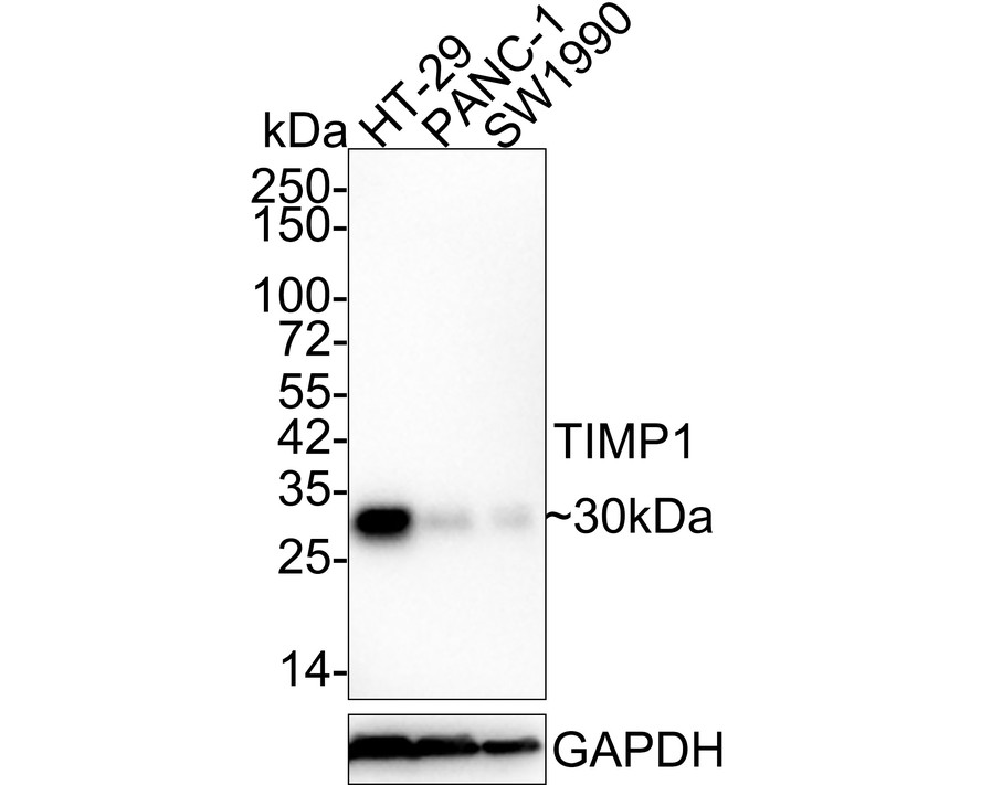
Western blot analysis of TIMP1 on different lysates with Rabbit anti-TIMP1 antibody at 1/1,000 dilution. Lane 1: HT-29 cell lysate, Lane 2: PANC-1 cell lysate, Lane 3: SW1990 cell lysate, Lysates/proteins at 40 µg/Lane. Exposure time: 3 minutes; 4-20% SDS-PAGE gel. Proteins were transferred to a PVDF membrane and blocked with 5% NFDM/TBST for 1 hour at room temperature. The primary antibody at 1/1,000 dilution was used in 5% NFDM/TBST at 4℃ overnight. Goat Anti-Rabbit IgG - HRP Secondary Antibody at 1/50,000 dilution was used for 1 hour at room temperature.IHC
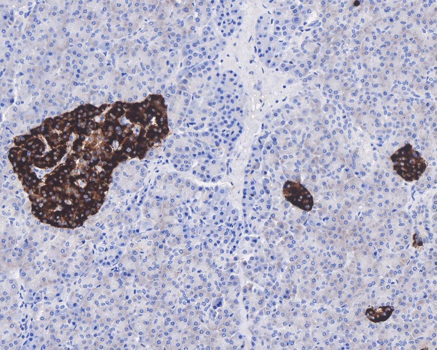
Immunohistochemical analysis of paraffin-embedded human pancreas tissue with Rabbit anti-TIMP1 antibody at 1/1,000 dilution. The section was pre-treated using heat mediated antigen retrieval with Tris-EDTA buffer (pH 9.0) for 20 minutes. The tissues were blocked in 1% BSA for 20 minutes at room temperature, washed with ddH2O and PBS, and then probed with the primary antibody at 1/1,000 dilution for 1 hour at room temperature. The detection was performed using an HRP conjugated compact polymer system. DAB was used as the chromogen. Tissues were counterstained with hematoxylin and mounted with DPX.FC
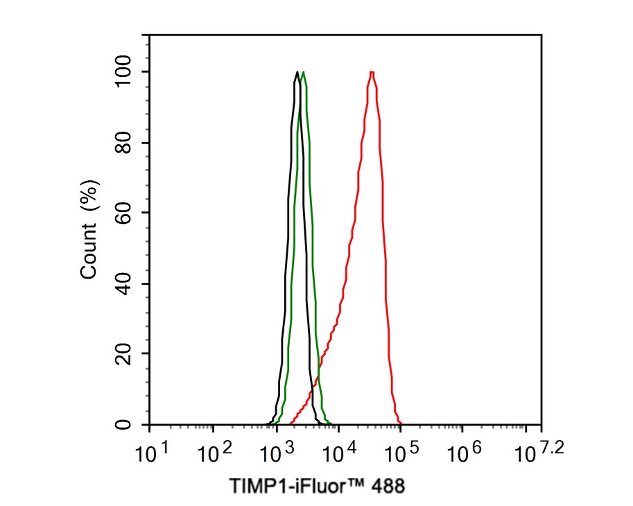
Flow cytometric analysis of HT-29 cells labeling TIMP1. Cells were fixed and permeabilized. Then stained with the primary antibody (1μg/mL) (red) compared with Rabbit IgG Isotype Control (green). After incubation of the primary antibody at +4℃ for an hour, the cells were stained with a 488 conjugate-Goat anti-Rabbit IgG Secondary antibody at 1/1,000 dilution for 30 minutes at +4℃. Unlabelled sample was used as a control (cells without incubation with primary antibody; black).| Product Name | TIMP1 Recombinant Rabbit Monoclonal Antibody |
|---|---|
| Antibody Type | Primary Antibodies |
| Immunogen | Recombinant full length protein within human TIMP1. |
| Clonality | monoclonal |
|---|---|
| Isotype | IgG |
| Host Species | Rabbit |
| Tested Applications | FCIHCWB |
| WB:1:1000 IHC:1:1000 FC:1:1000 |
|
| Species Reactivity | Human |
| Concentration | 1mg/ml |
| Purification | Protein A |
| Gene Symbol | TIMP1 |
|---|---|
| Gene Synonyms | EPA EPO HCI CLGI TIMP TIMP-1 |
| Gene Full Name | TIMP metallopeptidase inhibitor 1 |
| Gene Summary | This gene belongs to the TIMP gene family. The proteins encoded by this gene family are natural inhibitors of the matrix metalloproteinases (MMPs), a group of peptidases involved in degradation of the extracellular matrix. In addition to its inhibitory role against most of the known MMPs, the encoded protein is able to promote cell proliferation in a wide range of cell types, and may also have an anti-apoptotic function. Transcription of this gene is highly inducible in response to many cytokines and hormones. In addition, the expression from some but not all inactive X chromosomes suggests that this gene inactivation is polymorphic in human females. This gene is located within intron 6 of the synapsin I gene and is transcribed in the opposite direction. [provided by RefSeq, Jul 2008] |
| Alternative Names | Clgi antibody Collagenase inhibitor antibody Collagenase inhibitor Human antibody EPA antibody EPO antibody Erythroid Potentiating Activity antibody Erythroid-potentiating activity antibody Fibroblast collagenase inhibitor antibody FLJ90373 antibody HCI antibody Clgi antibody Collagenase inhibitor antibody Collagenase inhibitor Human antibody EPA antibody EPO antibody Erythroid Potentiating Activity antibody Erythroid-potentiating activity antibody Fibroblast collagenase inhibitor antibody FLJ90373 antibody HCI antibody Human Collagenase Inhibitor antibody Metalloproteinase inhibitor 1 antibody Metalloproteinase inhibitor 1 precursor antibody OTTHUMP00000023214 antibody TIMP 1 antibody TIMP antibody TIMP metallopeptidase inhibitor 1 antibody TIMP-1 antibody Timp1 antibody TIMP1 protein antibody TIMP1_HUMAN antibody Tissue Inhibitor of Metalloproteinase 1 antibody Tissue inhibitor of metalloproteinases 1 antibody Tissue inhibitor of metalloproteinases antibody Ttissue inhibitor of metalloproteinase 1 erythroid potentiating activity collagenase inhibitor antibody |
| Molecular Weight(MW) | 23kDa(Observed band size: 30kDa) |
| Cellular Localization | Secreted. |
WB
Western blot analysis of TIMP1 on different lysates with Rabbit anti-TIMP1 antibody at 1/1,000 dilution. Lane 1: HT-29 cell lysate, Lane 2: PANC-1 cell lysate, Lane 3: SW1990 cell lysate, Lysates/proteins at 40 µg/Lane. Exposure time: 3 minutes; 4-20% SDS-PAGE gel. Proteins were transferred to a PVDF membrane and blocked with 5% NFDM/TBST for 1 hour at room temperature. The primary antibody at 1/1,000 dilution was used in 5% NFDM/TBST at 4℃ overnight. Goat Anti-Rabbit IgG - HRP Secondary Antibody at 1/50,000 dilution was used for 1 hour at room temperature.
IHC
Immunohistochemical analysis of paraffin-embedded human pancreas tissue with Rabbit anti-TIMP1 antibody at 1/1,000 dilution. The section was pre-treated using heat mediated antigen retrieval with Tris-EDTA buffer (pH 9.0) for 20 minutes. The tissues were blocked in 1% BSA for 20 minutes at room temperature, washed with ddH2O and PBS, and then probed with the primary antibody at 1/1,000 dilution for 1 hour at room temperature. The detection was performed using an HRP conjugated compact polymer system. DAB was used as the chromogen. Tissues were counterstained with hematoxylin and mounted with DPX.
FC
Flow cytometric analysis of HT-29 cells labeling TIMP1. Cells were fixed and permeabilized. Then stained with the primary antibody (1μg/mL) (red) compared with Rabbit IgG Isotype Control (green). After incubation of the primary antibody at +4℃ for an hour, the cells were stained with a 488 conjugate-Goat anti-Rabbit IgG Secondary antibody at 1/1,000 dilution for 30 minutes at +4℃. Unlabelled sample was used as a control (cells without incubation with primary antibody; black).| Application Notes | WB:1:1000 IHC:1:1000 FC:1:1000 |
|---|
| Form | Liquid |
|---|---|
| Storage Instructions | Store at +4℃ after thawing. Aliquot store at -20℃. Avoid repeated freeze / thaw cycles. |
| Storage Buffer | 1*TBS (pH7.4), 0.05% BSA, 40% Glycerol. Preservative: 0.05% Sodium Azide. |


