Application
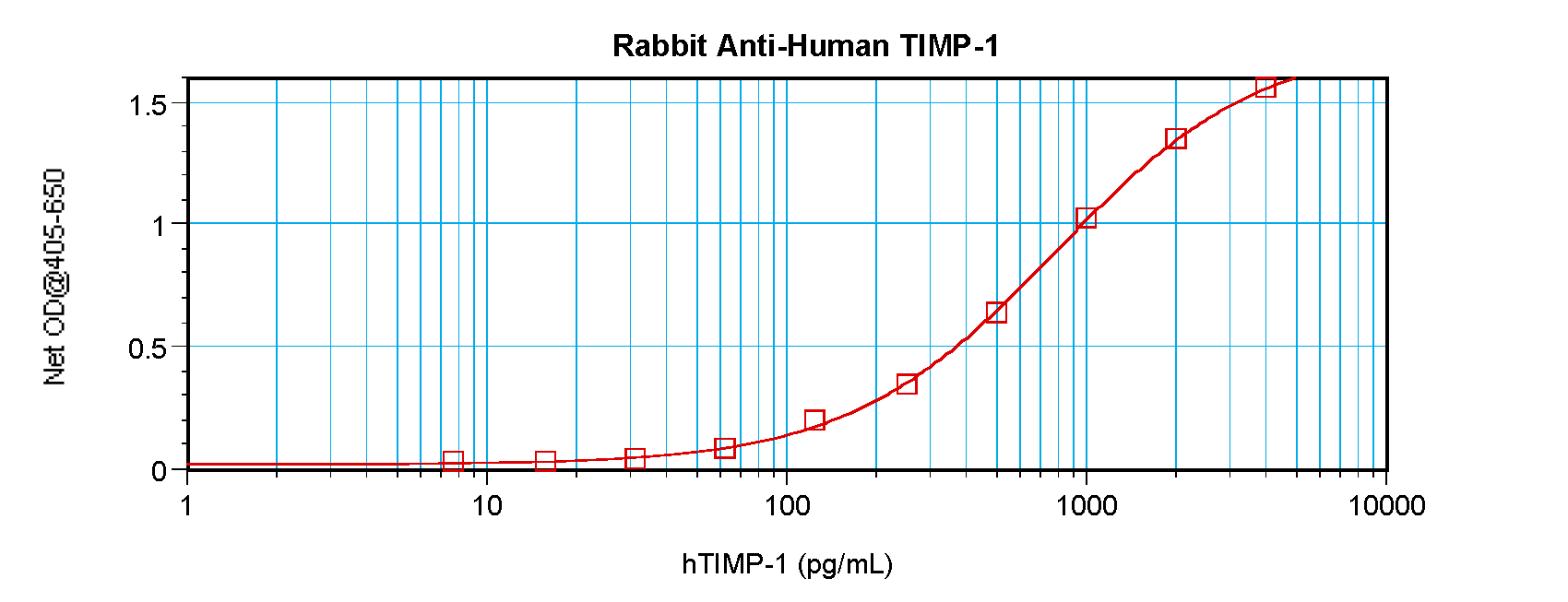
To detect hTIMP-1 by sandwich ELISA (using 100ul/well antibody solution) a concentration of 0.5 - 2.0 ug/ml of this antibody is required. This antigen affinity purified antibody, in conjunction with ProSci’s Biotinylated Anti-Human TIMP-1 (38-216) as a detection antibody, allows the detection of at least 0.2 - 0.4 ng/well of recombinant hTIMP-1.Application
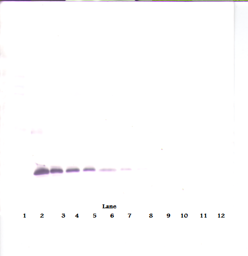
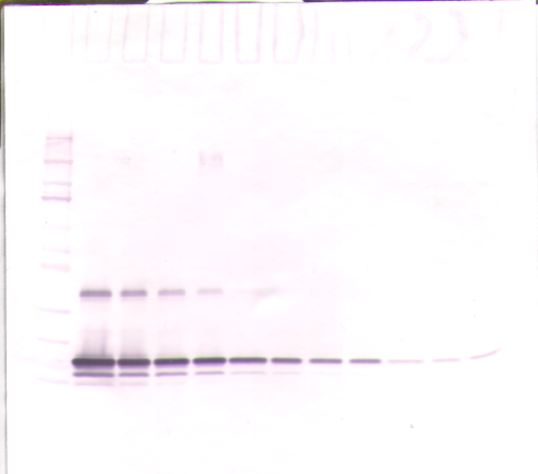
To detect hTIMP-1 by Western Blot analysis this antibody can be used at a concentration of 0.1 - 0.2 ug/ml. Used in conjunction with compatible secondary reagents the detection limit for recombinant hTIMP-1 is 1.5 - 3.0 ng/lane, under either reducing or non-reducing conditions.Application
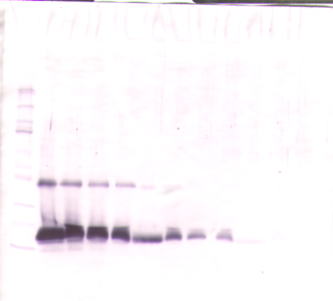
To detect hTIMP-1 by Western Blot analysis this antibody can be used at a concentration of 0.1 - 0.2 ug/ml. Used in conjunction with compatible secondary reagents the detection limit for recombinant hTIMP-1 is 1.5 - 3.0 ng/lane, under either reducing or non-reducing conditions.Application
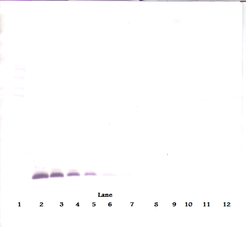
To detect hTIMP-1 by Western Blot analysis this antibody can be used at a concentration of 0.1 - 0.2 ug/ml. Used in conjunction with compatible secondary reagents the detection limit for recombinant hTIMP-1 is 1.5 - 3.0 ng/lane, under either reducing or non-reducing conditions.Application
To detect hTIMP-1 by Western Blot analysis this antibody can be used at a concentration of 0.1 - 0.2 ug/ml. Used in conjunction with compatible secondary reagents the detection limit for recombinant hTIMP-1 is 1.5 - 3.0 ng/lane, under either reducing or non-reducing conditions.Application
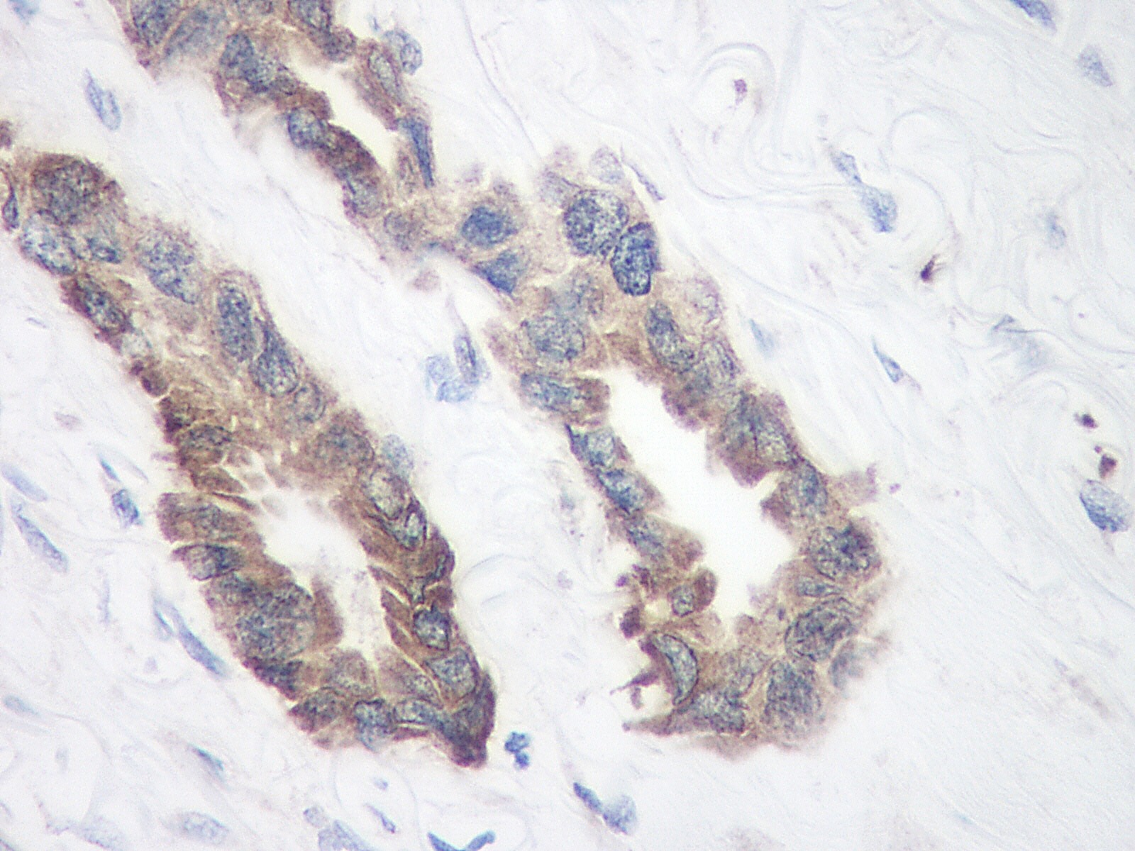
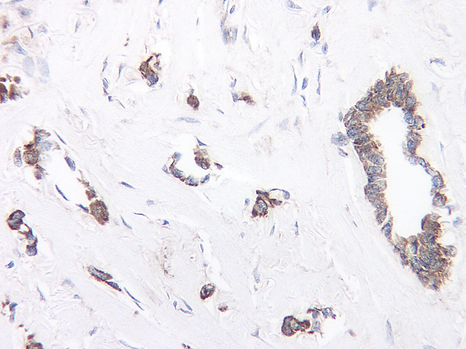
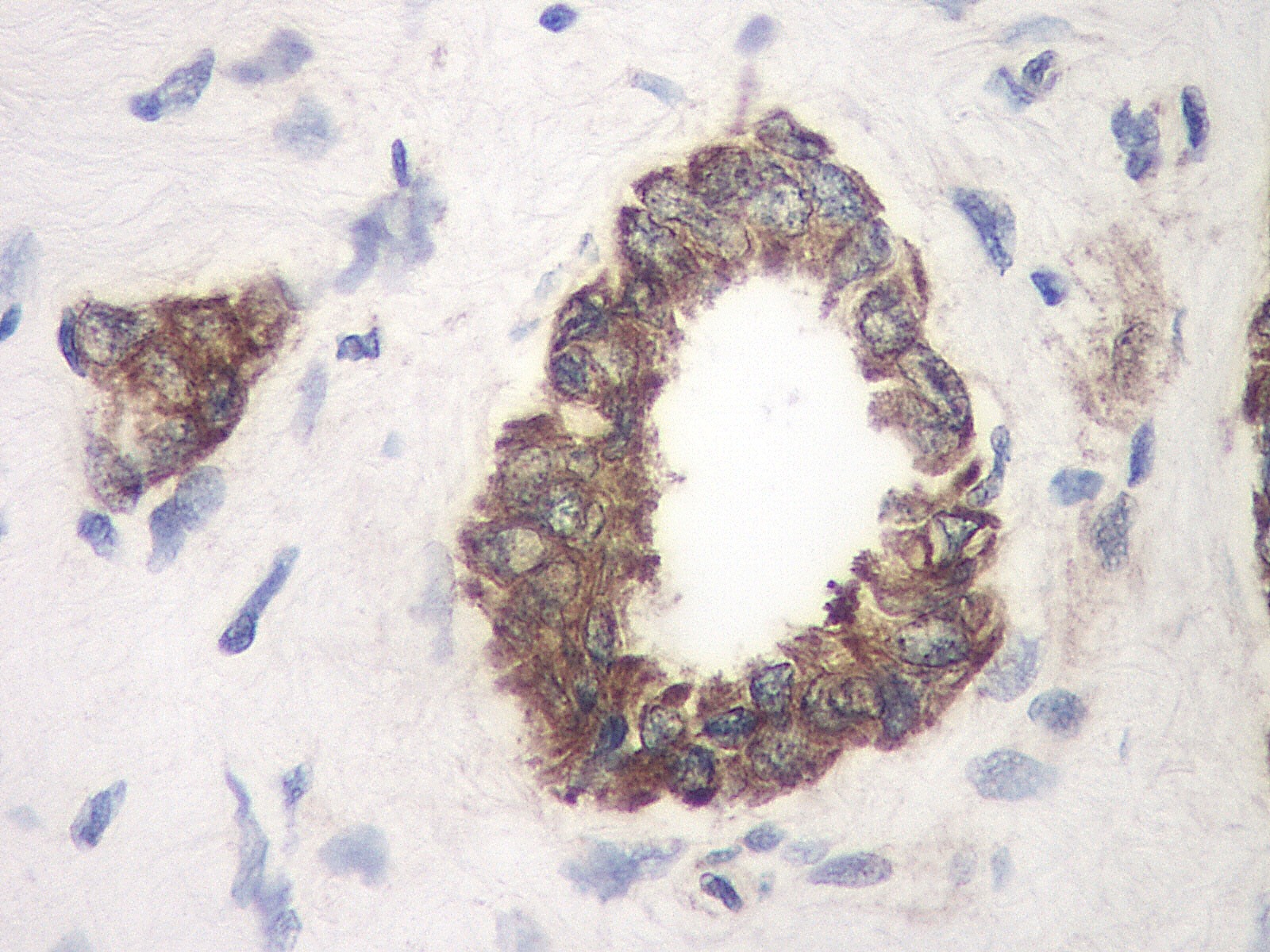
This antibody stained formalin-fixed, paraffin-embedded sections of human breast invasive ductal carcinoma. The recommended concentration is 0.1ug/ml with an overnight incubation at 4˚C. An HRP-labeled polymer detection system was used with a DAB chromogen. Optimal results were achieved with a proteinase K antigen retrieval. Optimal concentrations and conditions may vary.Application
This antibody stained formalin-fixed, paraffin-embedded sections of human breast invasive ductal carcinoma. The recommended concentration is 0.1ug/ml with an overnight incubation at 4˚C. An HRP-labeled polymer detection system was used with a DAB chromogen. Optimal results were achieved with a proteinase K antigen retrieval. Optimal concentrations and conditions may vary.Application
This antibody stained formalin-fixed, paraffin-embedded sections of human breast invasive ductal carcinoma. The recommended concentration is 0.1ug/ml with an overnight incubation at 4˚C. An HRP-labeled polymer detection system was used with a DAB chromogen. Optimal results were achieved with a proteinase K antigen retrieval. Optimal concentrations and conditions may vary.| Product Name | TIMP-1 Antibody |
|---|---|
| Antibody Type | Primary Antibodies |
| Antigen Alias | EPA, EPO, HCI, CLGI, TIMP, Metalloproteinase inhibitor 1, Erythroid-potentiating activity, EPA |
| Product description | Tissue inhibitor of metalloproteinases 1 (TIMP1) and Tissue inhibitor of metalloproteinases 2 (TIMP 2) have similar properties, specifically in inhibiting enzymes of the matrix metalloproteinase family, and are thought to be of great importance in the maintenance of connective tissue integrity. TIMP1 forms a complex of 1:1 stoichiometry with activated interstitial collagenases, activated stromelysin, active form of 72kDa Type IV collagenase (also known as MMP2 or gelatinase A), and latent and active forms of 92kDa Type IV collagenase (also known as MMP0 or gelatinase B). TIMPs inhibit the proteolytic invasiveness of tumour cells and normal placental trophoblast cells. TIMP1 is produced in low (pg/mL) levels by most cell types. Treatment of cells with the phorbol ester TPA stimulates production of TIMP1 in some cell types, but the low protein levels produced often require concentration of cell culture media to visualize the bands by Western Blotting. |
| Immunogen | Produced from sera of rabbits pre-immunized with highly pure (>98%) recombinant hTIMP-1 (human Tissue Inhibitor of Metalloproteinases-1). Human TIMP-1 specific antibody was purified by affinity chromatography employing immobilized hTIMP-1 matrix. |
| Clonality | Polyclonal |
|---|---|
| Host Species | Rabbit |
| Tested Applications | ELISANeutWB |
| Neutralization: To yield one-half maximal inhibition [ND50] of the biological activity of Human TIMP-1 (3.0 μg/mL), a concentration of 5.0 μg/mL of this antibody is required. ELISA: Indirect: To detect hTIMP-1 by indirect ELISA (using 100 μL/well antibody solution) a concentration of 0.5 - 2.0 μg/mL of this antibody is required. This antigen affinity purified antibody, in conjunction with compatible secondary reagents, allows the detection of at least 0.2 - 0.4 ng/well of recombinant hTIMP-1. Sandwich To detect hTIMP-1 by sandwich ELISA (using 100 μL/well antibody solution) a concentration of 0.5 - 2.0 μg/mL of this antibody is required. This antigen affinity purified antibody, in conjunction with our biotinylated Anti-Human TIMP-1 as a detection antibody, allows the detection of at least 0.2 - 0.4 ng/well of recombinant hTIMP-1. Western Blot: To detect hTIMP-1 by Western Blot analysis this antibody can be used at a concentration of 0.1 - 0.2 μg/mL. Used in conjunction with compatible secondary reagents the detection limit for recombinant hTIMP-1 is 1.5 - 3.0 ng/lane, under either reducing or non-reducing conditions.: |
|
| Species Reactivity | Human |
| Concentration | 1mg/ml |
| Gene Symbol | TIMP1 |
|---|---|
| Alternative Names | EPA EPO HCI CLGI TIMP Metalloproteinase inhibitor 1 Erythroid-potentiating activity EPA |
Application
To detect hTIMP-1 by sandwich ELISA (using 100ul/well antibody solution) a concentration of 0.5 - 2.0 ug/ml of this antibody is required. This antigen affinity purified antibody, in conjunction with ProSci’s Biotinylated Anti-Human TIMP-1 (38-216) as a detection antibody, allows the detection of at least 0.2 - 0.4 ng/well of recombinant hTIMP-1.
Application
To detect hTIMP-1 by Western Blot analysis this antibody can be used at a concentration of 0.1 - 0.2 ug/ml. Used in conjunction with compatible secondary reagents the detection limit for recombinant hTIMP-1 is 1.5 - 3.0 ng/lane, under either reducing or non-reducing conditions.
Application
To detect hTIMP-1 by Western Blot analysis this antibody can be used at a concentration of 0.1 - 0.2 ug/ml. Used in conjunction with compatible secondary reagents the detection limit for recombinant hTIMP-1 is 1.5 - 3.0 ng/lane, under either reducing or non-reducing conditions.
Application
To detect hTIMP-1 by Western Blot analysis this antibody can be used at a concentration of 0.1 - 0.2 ug/ml. Used in conjunction with compatible secondary reagents the detection limit for recombinant hTIMP-1 is 1.5 - 3.0 ng/lane, under either reducing or non-reducing conditions.
Application
To detect hTIMP-1 by Western Blot analysis this antibody can be used at a concentration of 0.1 - 0.2 ug/ml. Used in conjunction with compatible secondary reagents the detection limit for recombinant hTIMP-1 is 1.5 - 3.0 ng/lane, under either reducing or non-reducing conditions.
Application
This antibody stained formalin-fixed, paraffin-embedded sections of human breast invasive ductal carcinoma. The recommended concentration is 0.1ug/ml with an overnight incubation at 4˚C. An HRP-labeled polymer detection system was used with a DAB chromogen. Optimal results were achieved with a proteinase K antigen retrieval. Optimal concentrations and conditions may vary.
Application
This antibody stained formalin-fixed, paraffin-embedded sections of human breast invasive ductal carcinoma. The recommended concentration is 0.1ug/ml with an overnight incubation at 4˚C. An HRP-labeled polymer detection system was used with a DAB chromogen. Optimal results were achieved with a proteinase K antigen retrieval. Optimal concentrations and conditions may vary.
Application
This antibody stained formalin-fixed, paraffin-embedded sections of human breast invasive ductal carcinoma. The recommended concentration is 0.1ug/ml with an overnight incubation at 4˚C. An HRP-labeled polymer detection system was used with a DAB chromogen. Optimal results were achieved with a proteinase K antigen retrieval. Optimal concentrations and conditions may vary.| Application Notes | Neutralization: To yield one-half maximal inhibition [ND50] of the biological activity of Human TIMP-1 (3.0 μg/mL), a concentration of 5.0 μg/mL of this antibody is required. ELISA: Indirect: To detect hTIMP-1 by indirect ELISA (using 100 μL/well antibody solution) a concentration of 0.5 - 2.0 μg/mL of this antibody is required. This antigen affinity purified antibody, in conjunction with compatible secondary reagents, allows the detection of at least 0.2 - 0.4 ng/well of recombinant hTIMP-1. Sandwich To detect hTIMP-1 by sandwich ELISA (using 100 μL/well antibody solution) a concentration of 0.5 - 2.0 μg/mL of this antibody is required. This antigen affinity purified antibody, in conjunction with our biotinylated Anti-Human TIMP-1 as a detection antibody, allows the detection of at least 0.2 - 0.4 ng/well of recombinant hTIMP-1. Western Blot: To detect hTIMP-1 by Western Blot analysis this antibody can be used at a concentration of 0.1 - 0.2 μg/mL. Used in conjunction with compatible secondary reagents the detection limit for recombinant hTIMP-1 is 1.5 - 3.0 ng/lane, under either reducing or non-reducing conditions.: |
|---|
| Form | Liquid |
|---|---|
| Storage Instructions | TIMP-1 antibody is stable for at least 2 years from date of receipt at -20˚C. The reconstituted antibody is stable for at least two weeks at 2-8˚C. Frozen aliquots are stable for at least 6 months when stored at -20˚C. Avoid repeated freeze-thaw cycles. |
| Storage Buffer | phosphate buffered saline , pH 7.4, 150mM NaCl, 0.02% sodium azide and 50% glycerol. |


