Application
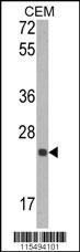
Western blot analysis of TIMP1 Antibody in CEM cell line lysates (35ug/lane)Application
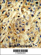
Formalin-fixed and paraffin-embedded human breast carcinoma reacted with TIMP1 Antibody (N-term), which was peroxidase-conjugated to the secondary antibody, followed by DAB staining.Application
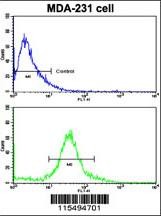
Flow cytometric analysis of MDA-231 cells using TIMP1 Antibody (N-term)(bottom histogram) compared to a negative control cell (top histogram). FITC-conjugated goat-anti-rabbit secondary antibodies were used for the analysis.Application
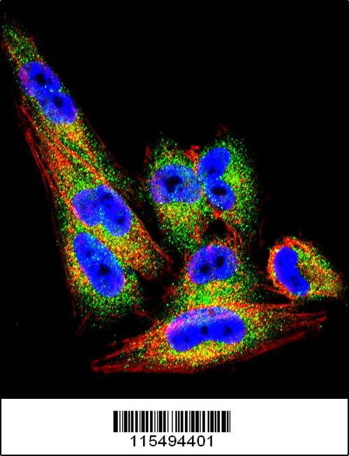
Confocal immunofluorescent analysis of TIMP1 Antibody with A2058 cell followed by Alexa Fluor 488-conjugated goat anti-rabbit lgG (green). Actin filaments have been labeled with Alexa Fluor 555 phalloidin (red).DAPI was used to stain the cell nuclear (blue).| Product Name | TIMP1 Antibody |
|---|---|
| Antibody Type | Primary Antibodies |
| Antigen Alias | Metalloproteinase inhibitor 1, Erythroid-potentiating activity, EPA, Fibroblast collagenase inhibitor, Collagenase inhibitor, Tissue inhibitor of metalloproteinases 1, TIMP-1, TIMP1, CLGI, TIMP |
| Product description | TIMP1 belongs to the TIMP family. The proteins in this family are natural inhibitors of the matrix metalloproteinases (MMPs), a group of peptidases involved in degradation of the extracellular matrix. In addition to its inhibitory role against most of the known MMPs, the protein is able to promote cell proliferation in a wide range of cell types, and may also have an anti-apoptotic function.1) Safranek,J., Anticancer Res. 29 (7), 2513-2517 (2009) Onal,I.K., Eur. J. Intern. Med. 20 (4), 369-372 (2009) Okamura,H., Int. J. Oncol. 35 (1), 181-186 (2009) |
| Immunogen | This TIMP1 antibody is generated from rabbits immunized with a KLH conjugated synthetic peptide between 41-70 amino acids from the N-terminal region of human TIMP1. |
| Modification | Acetylated |
| Clonality | Polyclonal |
|---|---|
| Isotype | Ig |
| Host Species | Rabbit |
| Tested Applications | FACSIFIHC-PWB |
| For WB starting dilution is: 1:1000 For IHC-P starting dilution is: 1:10~50 For FACS starting dilution is: 1:10~50 For IF starting dilution is: 1:10~50: |
|
| Species Reactivity | HumanMouseRat |
| Concentration | 1mg/ml |
| Gene Symbol | TIMP1 |
|---|---|
| Alternative Names | Metalloproteinase inhibitor 1 Erythroid-potentiating activity EPA Fibroblast collagenase inhibitor Collagenase inhibitor Tissue inhibitor of metalloproteinases 1 TIMP-1 TIMP1 CLGI TIMP |
| Molecular Weight(MW) | 23 kDa |
Application
Western blot analysis of TIMP1 Antibody in CEM cell line lysates (35ug/lane)
Application
Formalin-fixed and paraffin-embedded human breast carcinoma reacted with TIMP1 Antibody (N-term), which was peroxidase-conjugated to the secondary antibody, followed by DAB staining.
Application
Flow cytometric analysis of MDA-231 cells using TIMP1 Antibody (N-term)(bottom histogram) compared to a negative control cell (top histogram). FITC-conjugated goat-anti-rabbit secondary antibodies were used for the analysis.
Application
Confocal immunofluorescent analysis of TIMP1 Antibody with A2058 cell followed by Alexa Fluor 488-conjugated goat anti-rabbit lgG (green). Actin filaments have been labeled with Alexa Fluor 555 phalloidin (red).DAPI was used to stain the cell nuclear (blue).| Application Notes | For WB starting dilution is: 1:1000 For IHC-P starting dilution is: 1:10~50 For FACS starting dilution is: 1:10~50 For IF starting dilution is: 1:10~50: |
|---|
| Form | Liquid |
|---|---|
| Storage Instructions | Store at 4˚C for three months and -20˚C, stable for up to one year. As with all antibodies care should be taken to avoid repeated freeze thaw cycles. Antibodies should not be exposed to prolonged high temperatures. |
| Storage Buffer | Supplied in PBS with 0.09% (W/V) sodium azide. |


