WB
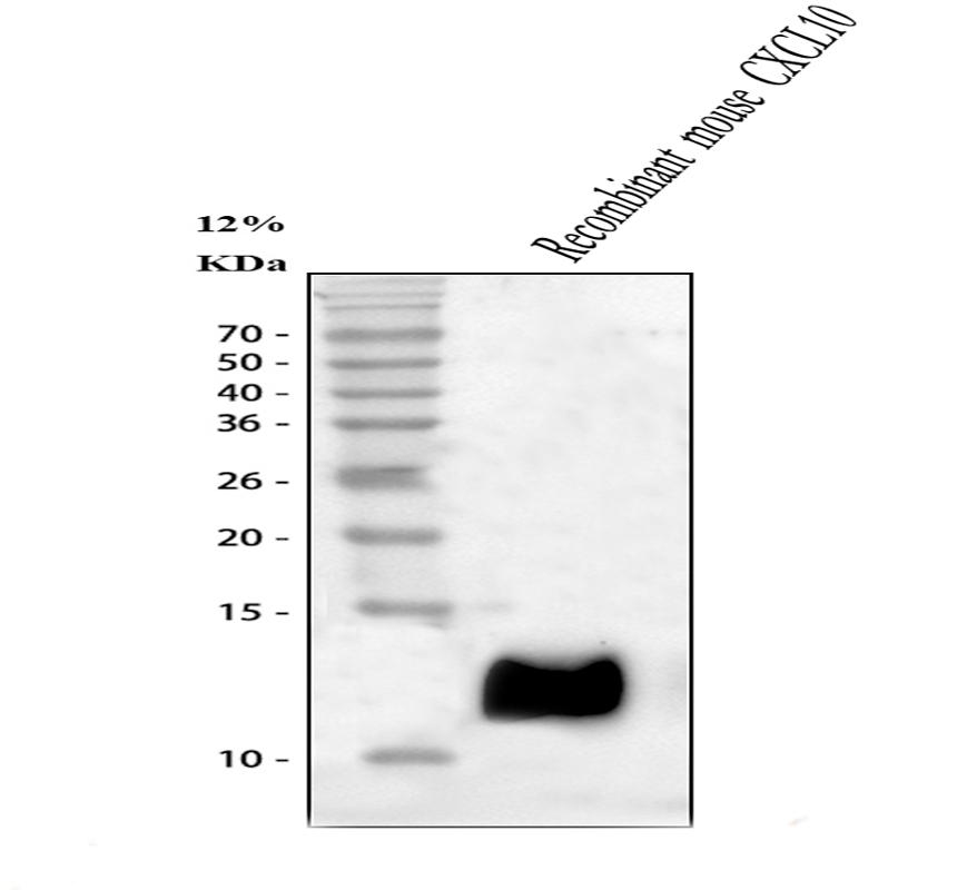
Western blot analysis of IP10/CXCL10 using anti-IP10/CXCL10 antibody. Lane 1: recombinant mouse CXCL10 protein 10ng. After electrophoresis, proteins were transferred to a membrane. Then the membrane was incubated with rabbit anti-IP10/CXCL10 antigen affinity purified polyclonal antibody at a dilution of 1:1000 and probed with a goat anti-rabbit IgG-HRP secondary antibody. The signal is developed using ECL Plus Western Blotting Substrate.IHC
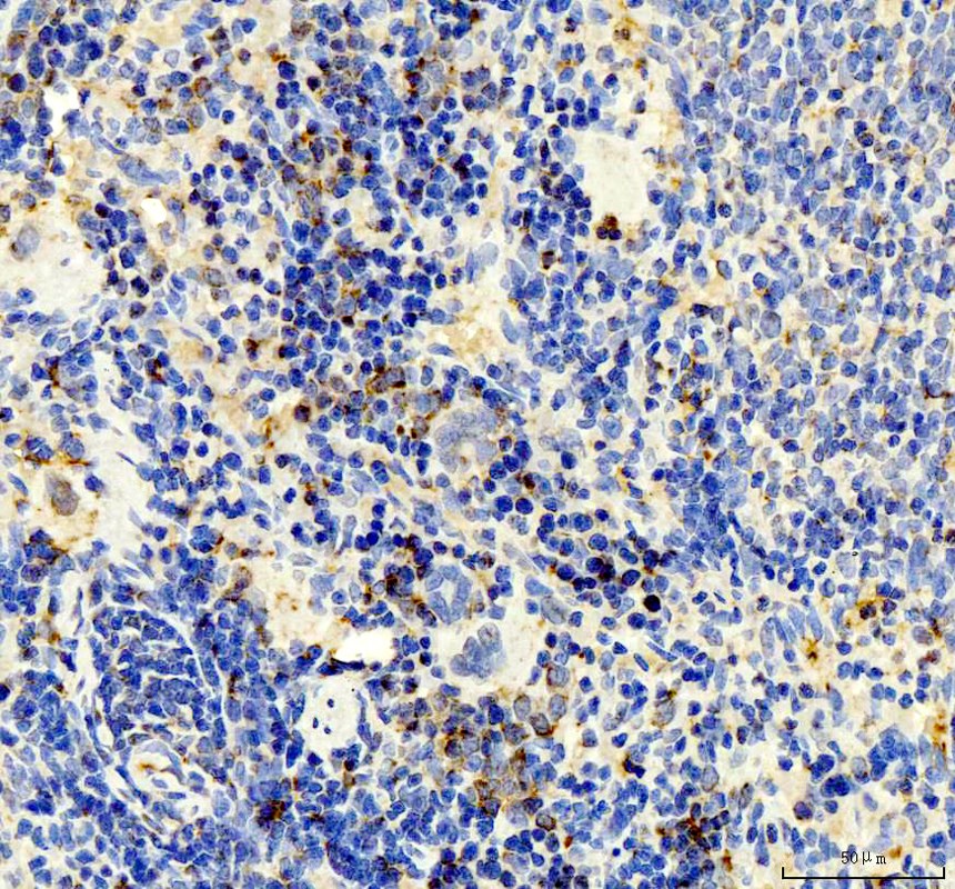
P10/CXCL10 was detected in a paraffin-embedded section of rat spleen tissue. The tissue section was incubated with rabbit anti-IP10/CXCL10 Antibody at a dilution of 1:200 and developed using HRP Conjugated Rabbit IgG Super Vision Assay Kit with DAB as the chromogen.IHC
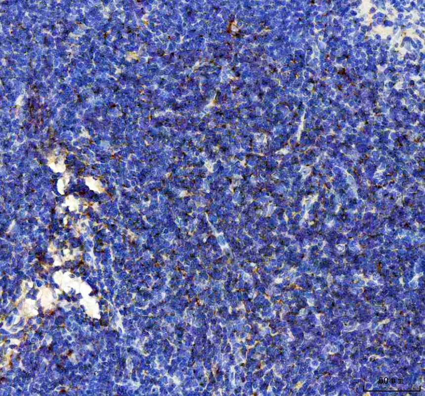
IP10/CXCL10 was detected in a paraffin-embedded section of mouse spleen tissue. The tissue section was incubated with rabbit anti-IP10/CXCL10 Antibody at a dilution of 1:200 and developed using HRP Conjugated Rabbit IgG Super Vision Assay Kit with DAB as the chromogen.| Product Name | Rabbit polyclonal antibody to IP10/CXCL10 |
|---|---|
| Antibody Type | Primary Antibodies |
| Immunogen | E. coli-derived mouse IP10 recombinant protein (Position: I22-P98). Mouse IP10 shares 70.1% and 77.9% amino acid (aa) sequence identity with human and rat IP10, respectively. |
| Clonality | Polyclonal |
|---|---|
| Isotype | IgG |
| Host Species | Rabbit |
| Tested Applications | IHCWB |
| WB:1:500-1:2000 IHC:1:50-1:400 |
|
| Species Reactivity | MouseRat |
| Concentration | 0.5mg/ml |
| Purification | Affinity purified |
| Gene Symbol | Cxcl10 |
|---|---|
| Gene Synonyms | C7 IP10 CRG-2 INP10 IP-10 Ifi10 mob-1 Scyb10 gIP-10 |
| Gene Full Name | C-X-C motif chemokine ligand 10 |
| Gene Summary | Predicted to enable chemoattractant activity; chemokine receptor binding activity; and heparin binding activity. Involved in cellular response to interleukin-17. Acts upstream of or within several processes, including defense response to virus; negative regulation of myoblast differentiation; and negative regulation of myoblast fusion. Located in external side of plasma membrane and extracellular space. Is expressed in several structures, including alimentary system; axial skeleton; hemolymphoid system; lacrimal gland; and liver. Human ortholog(s) of this gene implicated in hepatitis B; middle cerebral artery infarction; and type 1 diabetes mellitus. Orthologous to human CXCL10 (C-X-C motif chemokine ligand 10). [provided by Alliance of Genome Resources, Jul 2025] |
| Alternative Names | C X C motif chemokine 10 C7 crg 2 CXCL10 CXCL10/IP10 Gamma IP10 gIP 10 IFI10 INP10 IP 10 IP10 mob 1 SCYB10 Small inducible cytokine B10 |
| Molecular Weight(MW) | 11 kDa |
| Cellular Localization | Secreted. |
WB
Western blot analysis of IP10/CXCL10 using anti-IP10/CXCL10 antibody. Lane 1: recombinant mouse CXCL10 protein 10ng. After electrophoresis, proteins were transferred to a membrane. Then the membrane was incubated with rabbit anti-IP10/CXCL10 antigen affinity purified polyclonal antibody at a dilution of 1:1000 and probed with a goat anti-rabbit IgG-HRP secondary antibody. The signal is developed using ECL Plus Western Blotting Substrate.
IHC
P10/CXCL10 was detected in a paraffin-embedded section of rat spleen tissue. The tissue section was incubated with rabbit anti-IP10/CXCL10 Antibody at a dilution of 1:200 and developed using HRP Conjugated Rabbit IgG Super Vision Assay Kit with DAB as the chromogen.
IHC
IP10/CXCL10 was detected in a paraffin-embedded section of mouse spleen tissue. The tissue section was incubated with rabbit anti-IP10/CXCL10 Antibody at a dilution of 1:200 and developed using HRP Conjugated Rabbit IgG Super Vision Assay Kit with DAB as the chromogen.| Application Notes | WB:1:500-1:2000 IHC:1:50-1:400 |
|---|
| Form | Liquid |
|---|---|
| Storage Instructions | 12 months from date of receipt, -20℃ as supplied. 6 months 2 to 8℃ after reconstitution. Avoid repeated freezing and thawing. |
| Storage Buffer | 500ug/ml antibody with PBS, 0.02% NaN3, 1 mg/ml BSA and 50% glycerol. |


