WB
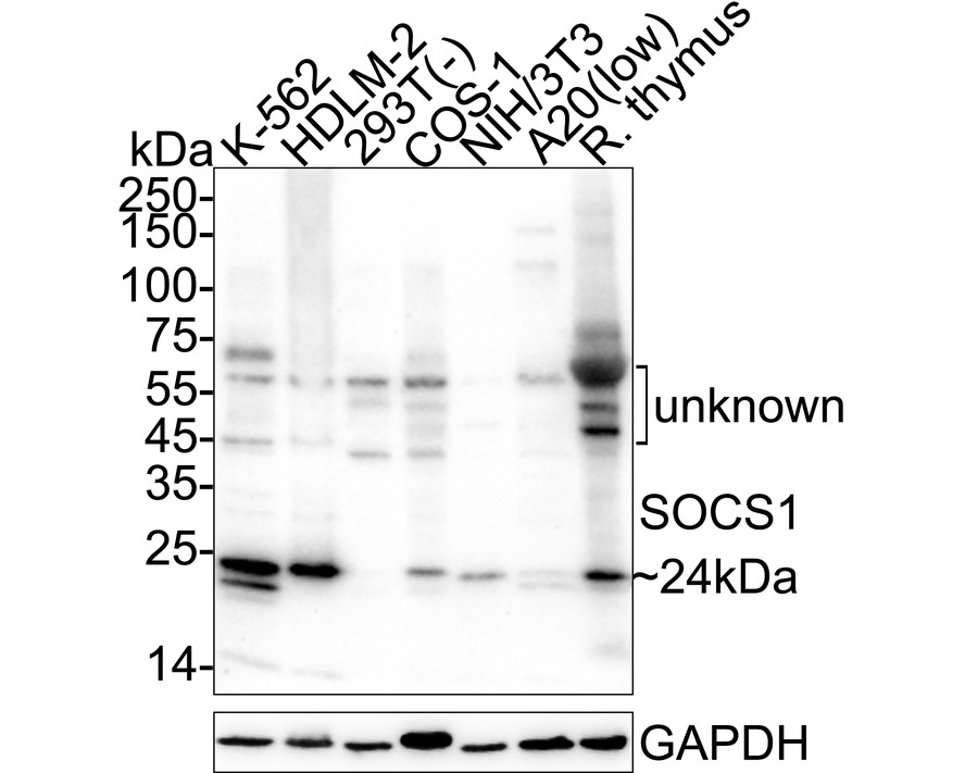
Western blot analysis of SOCS1 on different lysates with Rabbit anti-SOCS1 antibody at 1/1,000 dilution. Lane 1: K-562 cell lysate (30 µg/Lane), Lane 2: HDLM-2 cell lysate (30 µg/Lane), Lane 3: 293T cell lysate (negative) (30 µg/Lane), Lane 4: COS-1 cell lysate (30 µg/Lane), Lane 5: NIH/3T3 cell lysate (30 µg/Lane), Lane 6: A20 cell lysate (low expression) (30 µg/Lane), Lane 7: Rat thymus tissue lysate (40 µg/Lane), Exposure time: 3 minutes; 4-20% SDS-PAGE gel. Proteins were transferred to a PVDF membrane and blocked with 5% NFDM/TBST for 1 hour at room temperature. The primary antibody at 1/1,000 dilution was used in 5% NFDM/TBST at 4℃ overnight. Goat Anti-Rabbit IgG - HRP Secondary Antibody at 1/50,000 dilution was used for 1 hour at room temperature.| Product Name | SOCS1 Recombinant Rabbit Monoclonal Antibody |
|---|---|
| Antibody Type | Primary Antibodies |
| Immunogen | Recombinant protein within human SOCS1 aa 1-211. |
| Clonality | Monoclonal |
|---|---|
| Isotype | IgG |
| Host Species | Rabbit |
| Tested Applications | WB |
| WB:1:1000 |
|
| Species Reactivity | HumanMonkeyMouseRat |
| Concentration | 1mg/ml |
| Purification | Protein A |
| Gene Symbol | SOCS1 |
|---|---|
| Gene Synonyms | JAB CIS1 SSI1 TIP3 CISH1 SSI-1 TIP-3 AISIMD SOCS-1 |
| Gene Full Name | suppressor of cytokine signaling 1 |
| Gene Summary | This gene encodes a member of the STAT-induced STAT inhibitor (SSI), also known as suppressor of cytokine signaling (SOCS), family. SSI family members are cytokine-inducible negative regulators of cytokine signaling. The expression of this gene can be induced by a subset of cytokines, including IL2, IL3 erythropoietin (EPO), CSF2/GM-CSF, and interferon (IFN)-gamma. The protein encoded by this gene functions downstream of cytokine receptors, and takes part in a negative feedback loop to attenuate cytokine signaling. Knockout studies in mice suggested the role of this gene as a modulator of IFN-gamma action, which is required for normal postnatal growth and survival. [provided by RefSeq, Jul 2008] |
| Alternative Names | CIS1 antibody CISH 1 antibody CISH1 antibody Cytokine inducible SH2 protein 1 antibody JAB antibody JAK binding protein antibody JAK-binding protein antibody Janus kinase binding protein antibody SOCS 1 antibody SOCS-1 antibody CIS1 antibody CISH 1 antibody CISH1 antibody Cytokine inducible SH2 protein 1 antibody JAB antibody JAK binding protein antibody JAK-binding protein antibody Janus kinase binding protein antibody SOCS 1 antibody SOCS-1 antibody Socs1 antibody SOCS1_HUMAN antibody SSI 1 antibody SSI-1 antibody SSI1 antibody STAT induced STAT inhibitor 1 antibody STAT-induced STAT inhibitor 1 antibody Suppressor of cytokine signaling 1 antibody Supressor of cytokine signalling 1 antibody TEC interacting protein 3 antibody Tec-interacting protein 3 antibody TIP 3 antibody TIP-3 antibody TIP3 antibody |
| Molecular Weight(MW) | 24 kDa |
| Cellular Localization | Nucleus, Cytoplasmic vesicle. |
WB
Western blot analysis of SOCS1 on different lysates with Rabbit anti-SOCS1 antibody at 1/1,000 dilution. Lane 1: K-562 cell lysate (30 µg/Lane), Lane 2: HDLM-2 cell lysate (30 µg/Lane), Lane 3: 293T cell lysate (negative) (30 µg/Lane), Lane 4: COS-1 cell lysate (30 µg/Lane), Lane 5: NIH/3T3 cell lysate (30 µg/Lane), Lane 6: A20 cell lysate (low expression) (30 µg/Lane), Lane 7: Rat thymus tissue lysate (40 µg/Lane), Exposure time: 3 minutes; 4-20% SDS-PAGE gel. Proteins were transferred to a PVDF membrane and blocked with 5% NFDM/TBST for 1 hour at room temperature. The primary antibody at 1/1,000 dilution was used in 5% NFDM/TBST at 4℃ overnight. Goat Anti-Rabbit IgG - HRP Secondary Antibody at 1/50,000 dilution was used for 1 hour at room temperature.| Application Notes | WB:1:1000 |
|---|
| Form | Liquid |
|---|---|
| Storage Instructions | Store at +4℃ after thawing. Aliquot store at -20℃. Avoid repeated freeze / thaw cycles. |
| Storage Buffer | PBS (pH7.4), 0.1% BSA, 40% Glycerol. Preservative: 0.05% Sodium Azide. |


