WB
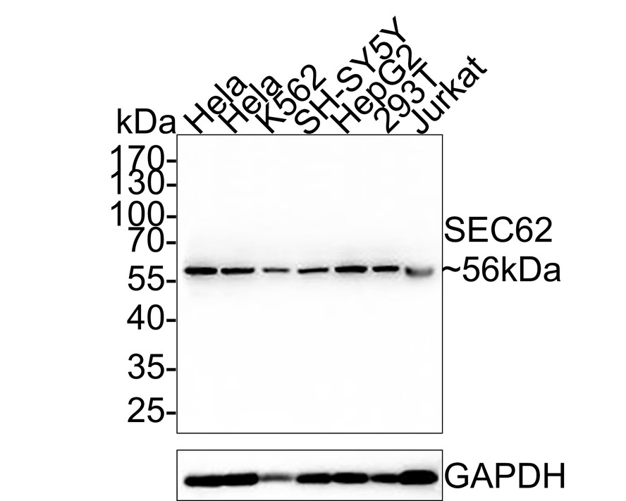
Western blot analysis of SEC62 on different lysates with Rabbit anti-SEC62 antibody at 1/5,000 dilution. Lane 1: Hela cell lysate, Lane 2: Hela cell lysate, Lane 3: K562 cell lysate, Lane 4: SH-SY5Y cell lysate, Lane 5: HepG2 cell lysate, Lane 6: 293T cell lysate, Lane 7: Jurkat cell lysate, Lysates/proteins at 20 µg/Lane. Exposure time: 30 seconds; 10% SDS-PAGE gel. Proteins were transferred to a PVDF membrane and blocked with 5% NFDM/TBST for 1 hour at room temperature. The primary antibody at 1/5,000 dilution was used in 5% NFDM/TBST at room temperature for 2 hours. Goat Anti-Rabbit IgG - HRP Secondary Antibody at 1:200,000 dilution was used for 1 hour at room temperature.WB
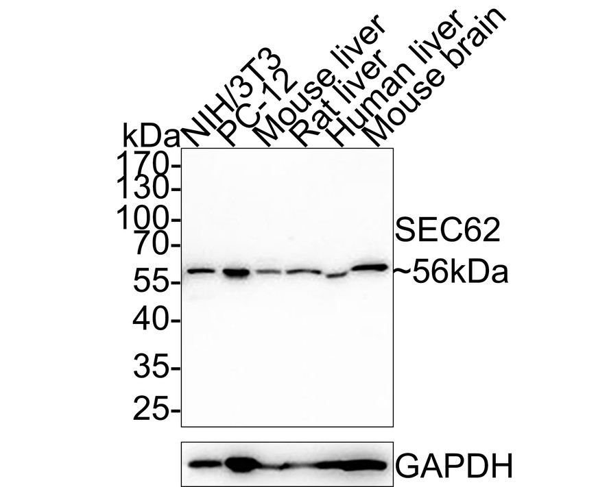
Western blot analysis of SEC62 on different lysates with Rabbit anti-SEC62 antibody at 1/5,000 dilution. Lane 1: NIH/3T3 cell lysate (20 µg/Lane), Lane 2: PC-12 cell lysate (20 µg/Lane), Lane 3: Mouse liver tissue lysate (40 µg/Lane), Lane 4: Rat liver tissue lysate (40 µg/Lane), Lane 5: Human liver tissue lysate (40 µg/Lane), Lane 6: Mouse brain tissue lysate (40 µg/Lane), Exposure time: 2 minutes; 10% SDS-PAGE gel. Proteins were transferred to a PVDF membrane and blocked with 5% NFDM/TBST for 1 hour at room temperature. The primary antibody at 1/5,000 dilution was used in 5% NFDM/TBST at room temperature for 2 hours. Goat Anti-Rabbit IgG - HRP Secondary Antibody at 1:200,000 dilution was used for 1 hour at room temperature.| Product Name | SEC62 Recombinant Rabbit Monoclonal Antibody |
|---|---|
| Antibody Type | Primary Antibodies |
| Immunogen | Synthetic peptide within human SEC62 aa 1-50 / 399. |
| Clonality | Monoclonal |
|---|---|
| Isotype | IgG |
| Host Species | Rabbit |
| Tested Applications | WB |
| WB:1:5000 |
|
| Species Reactivity | HumanMouseRat |
| Concentration | 1mg/ml |
| Purification | Protein A |
| Gene Symbol | SEC62 |
|---|---|
| Gene Synonyms | HTP1 TP-1 Dtrp1 TLOC1 |
| Gene Full Name | SEC62 preprotein translocation factor |
| Gene Summary | The Sec61 complex is the central component of the protein translocation apparatus of the endoplasmic reticulum (ER) membrane. The protein encoded by this gene and SEC63 protein are found to be associated with ribosome-free SEC61 complex. It is speculated that Sec61-Sec62-Sec63 may perform post-translational protein translocation into the ER. The Sec61-Sec62-Sec63 complex might also perform the backward transport of ER proteins that are subject to the ubiquitin-proteasome-dependent degradation pathway. The encoded protein is an integral membrane protein located in the rough ER. [provided by RefSeq, Jul 2008] |
| Alternative Names | Dtrp1 antibody FLJ32803 antibody hTP-1 antibody HTP1 antibody Membrane protein SEC62 S.cerevisiae homolog of antibody OTTHUMP00000213390 antibody sec62 antibody SEC62 homolog (S. cerevisiae) antibody SEC62_HUMAN antibody TLOC1 antibody Dtrp1 antibody FLJ32803 antibody hTP-1 antibody HTP1 antibody Membrane protein SEC62 S.cerevisiae homolog of antibody OTTHUMP00000213390 antibody sec62 antibody SEC62 homolog (S. cerevisiae) antibody SEC62_HUMAN antibody TLOC1 antibody TP-1 antibody Translocation protein 1 antibody Translocation protein sec62 antibody |
| Molecular Weight(MW) | 46 kDa(Observed band size:56 kDa) |
| Cellular Localization | Endoplasmic reticulum membrane. |
WB
Western blot analysis of SEC62 on different lysates with Rabbit anti-SEC62 antibody at 1/5,000 dilution. Lane 1: Hela cell lysate, Lane 2: Hela cell lysate, Lane 3: K562 cell lysate, Lane 4: SH-SY5Y cell lysate, Lane 5: HepG2 cell lysate, Lane 6: 293T cell lysate, Lane 7: Jurkat cell lysate, Lysates/proteins at 20 µg/Lane. Exposure time: 30 seconds; 10% SDS-PAGE gel. Proteins were transferred to a PVDF membrane and blocked with 5% NFDM/TBST for 1 hour at room temperature. The primary antibody at 1/5,000 dilution was used in 5% NFDM/TBST at room temperature for 2 hours. Goat Anti-Rabbit IgG - HRP Secondary Antibody at 1:200,000 dilution was used for 1 hour at room temperature.
WB
Western blot analysis of SEC62 on different lysates with Rabbit anti-SEC62 antibody at 1/5,000 dilution. Lane 1: NIH/3T3 cell lysate (20 µg/Lane), Lane 2: PC-12 cell lysate (20 µg/Lane), Lane 3: Mouse liver tissue lysate (40 µg/Lane), Lane 4: Rat liver tissue lysate (40 µg/Lane), Lane 5: Human liver tissue lysate (40 µg/Lane), Lane 6: Mouse brain tissue lysate (40 µg/Lane), Exposure time: 2 minutes; 10% SDS-PAGE gel. Proteins were transferred to a PVDF membrane and blocked with 5% NFDM/TBST for 1 hour at room temperature. The primary antibody at 1/5,000 dilution was used in 5% NFDM/TBST at room temperature for 2 hours. Goat Anti-Rabbit IgG - HRP Secondary Antibody at 1:200,000 dilution was used for 1 hour at room temperature.| Application Notes | WB:1:5000 |
|---|
| Form | Liquid |
|---|---|
| Storage Instructions | Store at +4℃ after thawing. Aliquot store at -20℃. Avoid repeated freeze / thaw cycles. |
| Storage Buffer | PBS (pH7.4), 0.1% BSA, 40% Glycerol. Preservative: 0.05% Sodium Azide. |


