WB
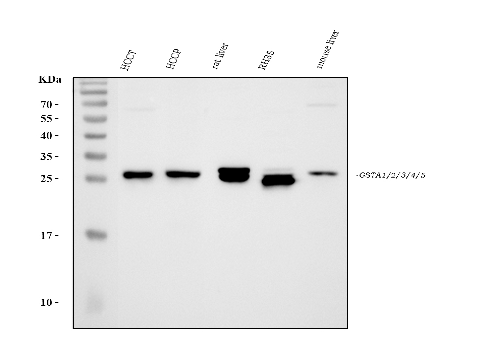
Western blot analysis of GSTA1/A2/A3/A4/A5 using anti-GSTA1/A2/A3/A4/A5 antibody. The sample well of each lane was loaded with 30 ug of sample under reducing conditions. Lane 1: HCCT whole cell lysates, Lane 2: HCCP whole cell lysates, Lane 3: rat liver tissue lysates, Lane 4: RH35 whole cell lysates, Lane 5: mouse liver tissue lysates. After electrophoresis, proteins were transferred to a membrane. Then the membrane was incubated with rabbit anti-GSTA1/A2/A3/A4/A5 antigen affinity purified polyclonal antibody at a dilution of 1:1000 and probed with a goat anti-rabbit IgG-HRP secondary antibody. The signal is developed using ECL Plus Western Blotting Substrate.IHC
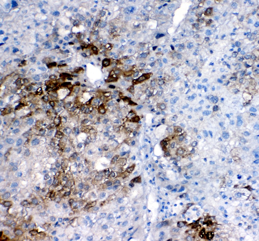
GSTA1/A2/A3/A4/A5 was detected in a paraffin-embedded section of human liver cancer tissue. The tissue section was incubated with rabbit anti-GSTA1/A2/A3/A4/A5 Antibody at a dilution of 1:200 and developed using HRP Conjugated Rabbit IgG Super Vision Assay Kit with DAB as the chromogen.IF-P
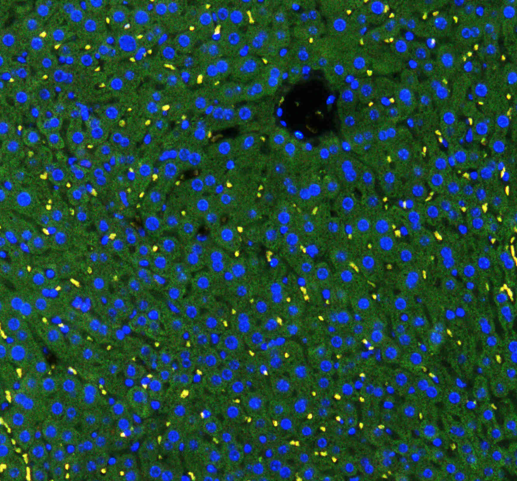
IF analysis using anti- GSTA1/A2/A3/A4/A5 antibody. detected in paraffin-embedded section of mouse liver tissue. The tissue section were stained using the Fluoro488-conjugated Anti-rabbit IgG Secondary Antibody (green) and counterstained with DAPI (blue).IF-P
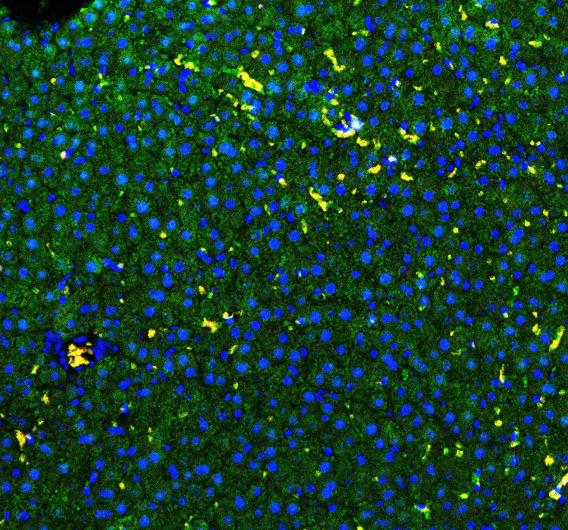
IF analysis using anti- GSTA1/A2/A3/A4/A5 antibody. detected in paraffin-embedded section of rat liver tissue. The tissue section were stained using the Fluoro488-conjugated Anti-rabbit IgG Secondary Antibody (green) and counterstained with DAPI (blue).| Product Name | Anti-GSTA1/A2/A3/A4/A5 Antibody |
|---|---|
| Antibody Type | Primary Antibodies |
| Product description | Cytosolic and membrane-bound forms of glutathione S-transferase are encoded by two distinct supergene families. These enzymes function in the detoxification of electrophilic compounds, including carcinogens, therapeutic drugs, environmental toxins and products of oxidative stress, by conjugation with glutathione. The genes encoding these enzymes are known to be highly polymorphic. These genetic variations can change an individual's susceptibility to carcinogens and toxins as well as affect the toxicity and efficacy of some drugs. At present, eight distinct classes of the soluble cytoplasmic mammalian glutathione S-transferases have been identified: alpha, kappa, mu, omega, pi, sigma, theta and zeta. This gene encodes a glutathione S-transferase belonging to the alpha class. The alpha class genes, located in a cluster mapped to chromosome 6, are the most abundantly expressed glutathione S-transferases in liver (hepatocytes) and kidney (proximal tubules). In addition to metabolizing bilirubin and certain anti-cancer drugs in the liver, the alpha class of these enzymes exhibit glutathione peroxidase activity, thereby protecting the cells from reactive oxygen species and the products of peroxidation. |
| Immunogen | A synthetic peptide corresponding to a sequence at the N-terminus of human GSTA1/A2/A3/A4/A5 different from the related mouse and rat sequences by five amino acids. |
| Clonality | Polyclonal |
|---|---|
| Isotype | IgG |
| Host Species | Rabbit |
| Tested Applications | IF-PIHCWB |
| WB:1:500-1:2000 IHC:1:50-1:400 IF-P:1:50-1:400 |
|
| Species Reactivity | HumanMouseRat |
| Concentration | 0.5mg/ml |
| Purification | Affinity purified |
| Alternative Names | Glutathione S-transferase A1 GST HA subunit 1 GST class-alpha member 1 GST-epsilon GSTA1-1 GTH1 Glutathione S-transferase A1 N-terminally processed GSTA1 GSTA1/A2/A3/A4/A5 |
|---|---|
| Molecular Weight(MW) | 26 kDa |
| Cellular Localization | Cytoplasm. |
WB
Western blot analysis of GSTA1/A2/A3/A4/A5 using anti-GSTA1/A2/A3/A4/A5 antibody. The sample well of each lane was loaded with 30 ug of sample under reducing conditions. Lane 1: HCCT whole cell lysates, Lane 2: HCCP whole cell lysates, Lane 3: rat liver tissue lysates, Lane 4: RH35 whole cell lysates, Lane 5: mouse liver tissue lysates. After electrophoresis, proteins were transferred to a membrane. Then the membrane was incubated with rabbit anti-GSTA1/A2/A3/A4/A5 antigen affinity purified polyclonal antibody at a dilution of 1:1000 and probed with a goat anti-rabbit IgG-HRP secondary antibody. The signal is developed using ECL Plus Western Blotting Substrate.
IHC
GSTA1/A2/A3/A4/A5 was detected in a paraffin-embedded section of human liver cancer tissue. The tissue section was incubated with rabbit anti-GSTA1/A2/A3/A4/A5 Antibody at a dilution of 1:200 and developed using HRP Conjugated Rabbit IgG Super Vision Assay Kit with DAB as the chromogen.
IF-P
IF analysis using anti- GSTA1/A2/A3/A4/A5 antibody. detected in paraffin-embedded section of mouse liver tissue. The tissue section were stained using the Fluoro488-conjugated Anti-rabbit IgG Secondary Antibody (green) and counterstained with DAPI (blue).
IF-P
IF analysis using anti- GSTA1/A2/A3/A4/A5 antibody. detected in paraffin-embedded section of rat liver tissue. The tissue section were stained using the Fluoro488-conjugated Anti-rabbit IgG Secondary Antibody (green) and counterstained with DAPI (blue).| Application Notes | WB:1:500-1:2000 IHC:1:50-1:400 IF-P:1:50-1:400 |
|---|
| Form | Liquid |
|---|---|
| Storage Instructions | 12 months from date of receipt, -20℃ as supplied. 6 months 2 to 8℃ after reconstitution. Avoid repeated freezing and thawing. |
| Storage Buffer | 500ug/ml antibody with PBS, 0.02% NaN3, 1 mg/ml BSA and 50% glycerol. |


