WB
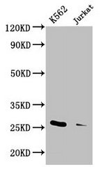
Positive WB detected in: K562 whole cell lysate, Jurkat whole cell lysate, All lanes: MOB1A antibody at 3.3µg/ml, Secondary Goat polyclonal to rabbit IgG at 1/50000 dilution.IHC
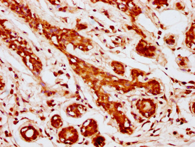
IHC image of MOB1A Antibody diluted at 1:1000 and staining in paraffin-embedded human breast cancer performed on a Leica BondTM system. After dewaxing and hydration, antigen retrieval was mediated by high pressure in a citrate buffer (pH 6.0). Section was blocked with 10% normal goat serum 30min at RT. Then primary antibody (1% BSA) was incubated at 4°C overnight. The primary is detected by a biotinylated secondary antibody and visualized using an HRP conjugated SP system.ICC/IF
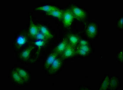
Immunofluorescence staining of HepG2 cells with MOB1A Antibody at 1:333, counter-stained with DAPI. The cells were fixed in 4% formaldehyde, permeabilized using 0.2% Triton X-100 and blocked in 10% normal Goat Serum. The cells were then incubated with the antibody overnight at 4°C. The secondary antibody was Alexa Fluor 488-congugated AffiniPure Goat Anti-Rabbit IgG(H+L).IP
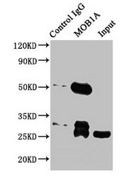
Immunoprecipitating MOB1A in Jurkat whole cell lysate, Lane 1: Rabbit control IgG instead of MOB1A Antibody in Jurkat whole cell lysate. For western blotting, a HRP-conjugated Protein G antibody was used as the secondary antibody (1/2000). Lane 2: MOB1A Antibody (5µg) + Jurkat whole cell lysate (500µg). Lane 3: Jurkat whole cell lysate (20µg).| Product Name | Rabbit anti-MOB1A Polyclonal antibody |
|---|---|
| Antibody Type | Primary Antibodies |
| Immunogen | Recombinant Human Neurexin-1-beta protein (184-363AA) |
| Clonality | Polyclonal |
|---|---|
| Isotype | IgG |
| Host Species | Rabbit |
| Tested Applications | ICC/IFIHCIPWB |
| WB:1:2000-1:5000 IHC:1:100-1:1000 ICC/IF:1:200-1:500 IP:1:200-1:2000 |
|
| Species Reactivity | Human |
| Concentration | 1mg/ml |
| Purification | Affinity purified |
| Gene Symbol | MOB1A |
|---|---|
| Gene Synonyms | MOB1 MATS1 Mob4B C2orf6 MOBK1B MABKL1B MOBKL1B |
| Gene Full Name | MOB kinase activator 1A |
| Gene Summary | The protein encoded by this gene is a component of the Hippo signaling pathway, which controls organ size and tumor growth by enhancing apoptosis. Loss of the encoded protein results in cell proliferation and cancer formation. The encoded protein is also involved in the control of microtubule stability during cytokinesis. Several transcript variants encoding different isoforms have been found for this gene. [provided by RefSeq, Nov 2015] |
| Alternative Names | B1 Mps One Binder kinase activator like 1B (yeast) antibody C2orf6 antibody Chromosome 2 open reading frame 6 antibody FLJ10788 antibody FLJ11595 antibody MATS1 antibody MOB kinase activator 1A antibody MOB1 antibody Mob1 alpha antibody Mob1 homolog 1B antibody MOB1 Mps One Binder homolog A antibody MOB1 Mps One Binder kinase activator like 1B antibody Mob1A antibody MOB4B antibody Mob4B protein antibody MOBK1B antibody Mobkl1b antibody MOL1B_HUMAN antibody Mps one binder kinase activator like 1B antibody Mps one binder kinase activator-like 1B antibody Protein Mob4B antibody |
| Molecular Weight(MW) | 26 kDa |
| Cellular Localization | cytoplasm,cytosol,nucleus. |
WB
Positive WB detected in: K562 whole cell lysate, Jurkat whole cell lysate, All lanes: MOB1A antibody at 3.3µg/ml, Secondary Goat polyclonal to rabbit IgG at 1/50000 dilution.
IHC
IHC image of MOB1A Antibody diluted at 1:1000 and staining in paraffin-embedded human breast cancer performed on a Leica BondTM system. After dewaxing and hydration, antigen retrieval was mediated by high pressure in a citrate buffer (pH 6.0). Section was blocked with 10% normal goat serum 30min at RT. Then primary antibody (1% BSA) was incubated at 4°C overnight. The primary is detected by a biotinylated secondary antibody and visualized using an HRP conjugated SP system.
ICC/IF
Immunofluorescence staining of HepG2 cells with MOB1A Antibody at 1:333, counter-stained with DAPI. The cells were fixed in 4% formaldehyde, permeabilized using 0.2% Triton X-100 and blocked in 10% normal Goat Serum. The cells were then incubated with the antibody overnight at 4°C. The secondary antibody was Alexa Fluor 488-congugated AffiniPure Goat Anti-Rabbit IgG(H+L).
IP
Immunoprecipitating MOB1A in Jurkat whole cell lysate, Lane 1: Rabbit control IgG instead of MOB1A Antibody in Jurkat whole cell lysate. For western blotting, a HRP-conjugated Protein G antibody was used as the secondary antibody (1/2000). Lane 2: MOB1A Antibody (5µg) + Jurkat whole cell lysate (500µg). Lane 3: Jurkat whole cell lysate (20µg).| Application Notes | WB:1:2000-1:5000 IHC:1:100-1:1000 ICC/IF:1:200-1:500 IP:1:200-1:2000 |
|---|
| Form | Liquid |
|---|---|
| Storage Instructions | Upon receipt, store at -20°C or -80°C. Avoid repeated freeze. |
| Storage Buffer | 0.03% Proclin 300 Constituents: 50% Glycerol, 0.01M PBS, PH 7.4 |


