WB
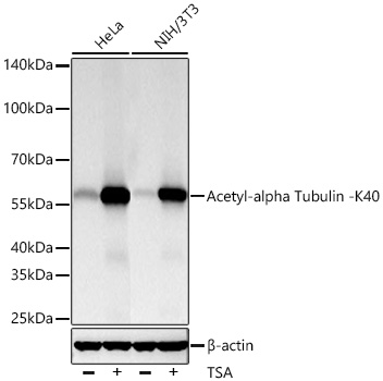
Western blot analysis of lysates from cells using Acetyl-alpha Tubulin -K40 Rabbit mAb at 1:10000 dilution incubated overnight at 4℃. HeLa cells and NIH/3T3 cells were treated with TSA (1 uM) at 37℃ for 18 hours. Secondary antibody: HRP-conjugated Goat anti-Rabbit IgG (H+L) at 1:10000 dilution. Lysates/proteins: 30 μg per lane. Blocking buffer: 3% nonfat dry milk in TBST. Detection: ECL Basic Kit. Exposure time: 10 s.IHC
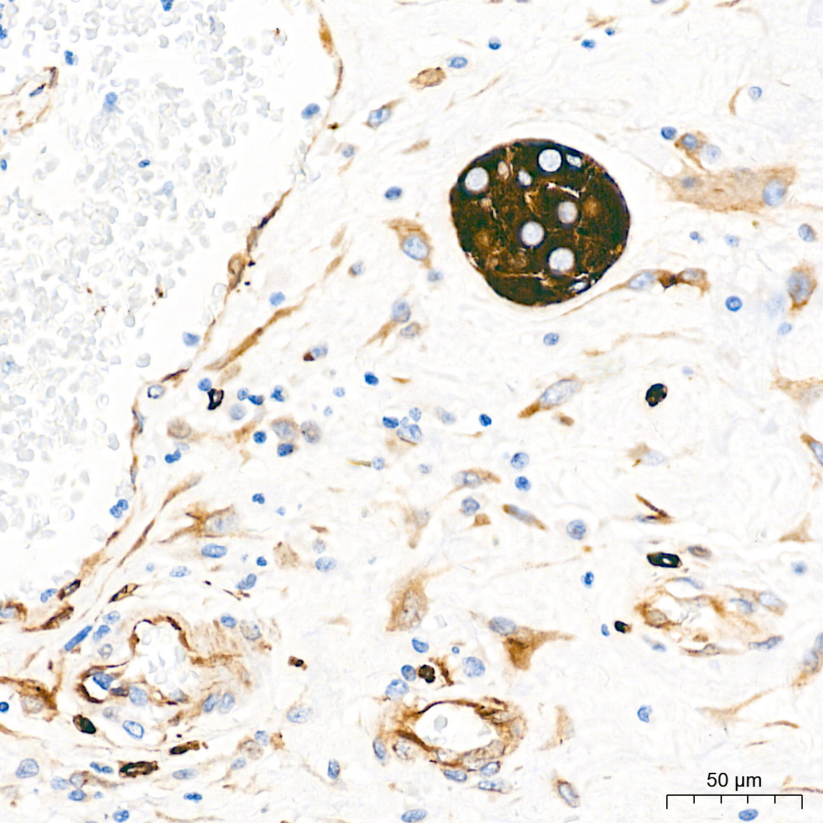
Immunohistochemistry analysis of paraffin embedded Human colon tissue using Acetyl alpha Tubulin -K40 Rabbit mAb at a dilution of 1:10000 (40x lens). High pressure antigen retrieval performed with 0.01M Tris EDTA Buffer (pH 9.0) prior to IHC staining.IHC
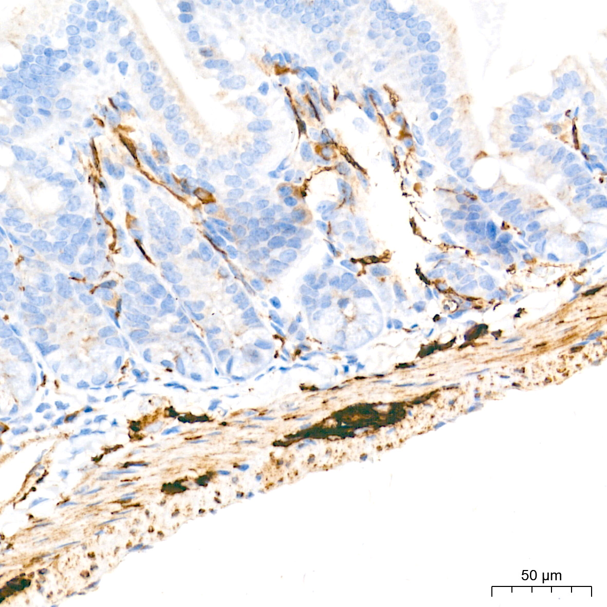
Immunohistochemistry analysis of paraffin embedded Moue intestin tissue using Acetyl alpha Tubulin -K40 Rabbit mAb at a dilution of 1:10000 (40x lens). High pressure antigen retrieval performed with 0.01M Tris EDTA Buffer (pH 9.0) prior to IHC staining.IHC
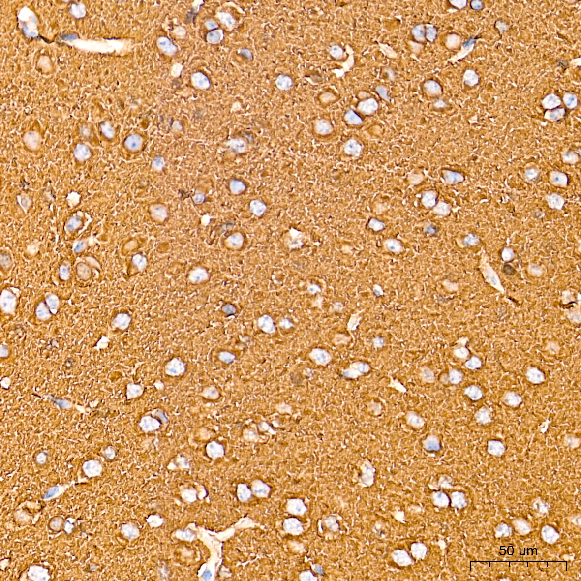
Immunohistochemistry analysis of paraffin embedded Rat brain tissue using Acetyl alpha Tubulin -K40 Rabbit mAb at a dilution of 1:10000 (40x lens). High pressure antigen retrieval performed with 0.01M Tris EDTA Buffer (pH 9.0) prior to IHC staining.ICC/IF
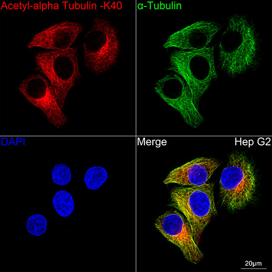
Confocal imaging of Hep G2 cells using Acetyl-alpha Tubulin -K40 Rabbit mAb (dilution 1:200) followed by a further incubation with Cy3 Goat Anti-Rabbit IgG (H+L) (dilution 1:500) (Red). The cells were counterstained with α-Tubulin mAb (dilution 1:400) followed by incubation with Omnimabs® 488 Goat Anti-Mouse IgG(H&L) Ab (dilution 1:500) (Green). DAPI was used for nuclear staining (blue). Objective: 100x.| Product Name | Acetyl-alpha Tubulin -K40 Rabbit mAb |
|---|---|
| Antibody Type | Primary Antibodies |
| Immunogen | Synthetic peptide. |
| Modification | Acetyl-K40 |
| Clonality | monoclonal |
|---|---|
| Isotype | IgG |
| Host Species | Rabbit |
| Tested Applications | ICC/IFIHCWB |
| WB:1:5000-1:40000 IHC:1:10000-1:40000 ICC/IF:1:50-1:200 |
|
| Species Reactivity | HumanMouseRat |
| Concentration | 1mg/ml |
| Purification | Affinity purified |
| Gene Symbol | TUBA1A |
|---|---|
| Gene Synonyms | LIS3 TUBA3 B-ALPHA-1 |
| Gene Full Name | tubulin alpha 1a |
| Gene Summary | Microtubules of the eukaryotic cytoskeleton perform essential and diverse functions and are composed of a heterodimer of alpha and beta tubulins. The genes encoding these microtubule constituents belong to the tubulin superfamily, which is composed of six distinct families. Genes from the alpha, beta and gamma tubulin families are found in all eukaryotes. The alpha and beta tubulins represent the major components of microtubules, while gamma tubulin plays a critical role in the nucleation of microtubule assembly. There are multiple alpha and beta tubulin genes, which are highly conserved among species. This gene encodes alpha tubulin and is highly similar to the mouse and rat Tuba1 genes. Northern blot studies have shown that the gene expression is predominantly found in morphologically differentiated neurologic cells. This gene is one of three alpha-tubulin genes in a cluster on chromosome 12q. Mutations in this gene cause lissencephaly type 3 (LIS3) - a neurological condition characterized by microcephaly, intellectual disability, and early-onset epilepsy caused by defective neuronal migration. Alternative splicing results in multiple transcript variants encoding distinct isoforms. [provided by RefSeq, Jul 2017] |
| Alternative Names | LIS3 TUBA3 B-ALPHA-1 Acetyl-alpha Tubulin -K40 |
| Molecular Weight(MW) | 55kDa |
| Cellular Localization | Cytoplasm, cytoskeleton. |
WB
Western blot analysis of lysates from cells using Acetyl-alpha Tubulin -K40 Rabbit mAb at 1:10000 dilution incubated overnight at 4℃. HeLa cells and NIH/3T3 cells were treated with TSA (1 uM) at 37℃ for 18 hours. Secondary antibody: HRP-conjugated Goat anti-Rabbit IgG (H+L) at 1:10000 dilution. Lysates/proteins: 30 μg per lane. Blocking buffer: 3% nonfat dry milk in TBST. Detection: ECL Basic Kit. Exposure time: 10 s.
IHC
Immunohistochemistry analysis of paraffin embedded Human colon tissue using Acetyl alpha Tubulin -K40 Rabbit mAb at a dilution of 1:10000 (40x lens). High pressure antigen retrieval performed with 0.01M Tris EDTA Buffer (pH 9.0) prior to IHC staining.
IHC
Immunohistochemistry analysis of paraffin embedded Moue intestin tissue using Acetyl alpha Tubulin -K40 Rabbit mAb at a dilution of 1:10000 (40x lens). High pressure antigen retrieval performed with 0.01M Tris EDTA Buffer (pH 9.0) prior to IHC staining.
IHC
Immunohistochemistry analysis of paraffin embedded Rat brain tissue using Acetyl alpha Tubulin -K40 Rabbit mAb at a dilution of 1:10000 (40x lens). High pressure antigen retrieval performed with 0.01M Tris EDTA Buffer (pH 9.0) prior to IHC staining.
ICC/IF
Confocal imaging of Hep G2 cells using Acetyl-alpha Tubulin -K40 Rabbit mAb (dilution 1:200) followed by a further incubation with Cy3 Goat Anti-Rabbit IgG (H+L) (dilution 1:500) (Red). The cells were counterstained with α-Tubulin mAb (dilution 1:400) followed by incubation with Omnimabs® 488 Goat Anti-Mouse IgG(H&L) Ab (dilution 1:500) (Green). DAPI was used for nuclear staining (blue). Objective: 100x.| Application Notes | WB:1:5000-1:40000 IHC:1:10000-1:40000 ICC/IF:1:50-1:200 |
|---|
| Form | Liquid |
|---|---|
| Storage Instructions | Store at -20℃. Avoid freeze / thaw cycles. |
| Storage Buffer | Buffer: PBS with 0.05% proclin300, 0.05% BSA, 50% glycerol, pH7.3. |


