WB
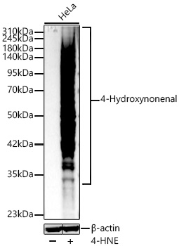
Western blot analysis of lysates from HeLa cells using 4-Hydroxynonenal Rabbit mAb at 1:10000 dilution incubated overnight at 4℃. HeLa cells were treated with 4-HNE (0.2 mg/ml) at 37℃ for 30 minutes. Secondary antibody: HRP-conjugated Goat anti-Rabbit IgG (H+L)at 1:10000 dilution. Lysates/proteins: 30 μg per lane. Blocking buffer: 3 % nonfat dry milk in TBST. Detection: ECL Basic Kit. Exposure time: 30s.ICC/IF
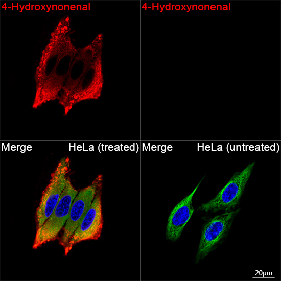
Confocal imaging of HeLa cells (treated with 4-HNE) and HeLa cells (untreated) using 4 Hydroxynonenal Rabbit mAb (dilution 1:200) followed by a further incubation with Cy3 Goat Anti-Rabbit IgG (H+L) (dilution 1:500) (Red). The cells were counterstained with α-Tubulin Mouse mAb (dilution 1:400) followed by incubation with Omnimabs® 488 Goat Anti-Mouse IgG(H&L) Ab (dilution 1:500) (Green). DAPI was used for nuclear staining (Blue). Objective: 100x.IF-P
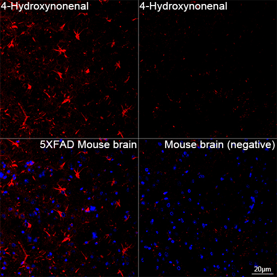
Confocal imaging of paraffin-embedded Mouse brain and 5xFAD Mouse brain tissue using 4-Hydroxynonenal Rabbit mAb (dilution 1:200) followed by a further incubation with Cy3 Goat Anti-Rabbit IgG (H+L) (dilution 1:500) (Red). DAPI was used for nuclear staining (Blue). High pressure antigen retrieval performed with 0.01M Citrate Buffer (pH 6.0) prior to IF staining. Objective: 40x.FC
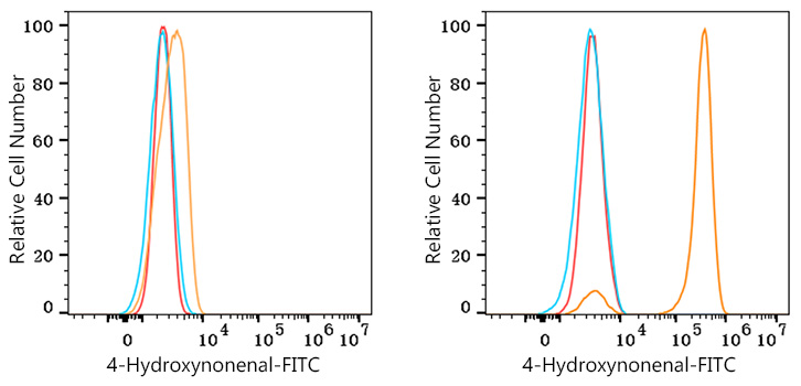
Flow cytometry: 1X10^6 HeLa cells (negative control,left) and HeLa cells (treated with 4-Hydroxynonenal,right) were intracellularly-stained with 4-Hydroxynonenal Rabbit mAb (2 μg/mL,orange line) or Rabbit IgG isotype control (2 μg/mL,blue line), followed by FITC conjugated goat anti-Rabbit pAb staining. Non-fluorescently stained cells were used as blank control (red line).| Product Name | 4-Hydroxynonenal Rabbit mAb |
|---|---|
| Antibody Type | Primary Antibodies |
| Product description | 4-hydroxy-2-nonenal (4-hydroxynonenal, 4-HNE) is a highly reactive aldehyde generated by the exposure of polyunsaturated fatty acids to peroxides and reactive oxygen species (ROS). It non-enzymatically forms stable protein adducts with histidine, lysine, and cysteine side chains that have been used as biomarkers for oxidative damage in cells. Conditions where 4-HNE immunoreactivity has been observed include include inflammation, neurodegenerative diseases, and ischemic damage to the heart and brain. |
| Immunogen | Chemical compounds corresponding to 4-Hydroxynonenal. |
| Clonality | monoclonal |
|---|---|
| Isotype | IgG |
| Host Species | Rabbit |
| Tested Applications | FCICC/IFIF-PWB |
| WB:1:1000-1:10000 ICC/IF:1:200-1:500 IF-P:1:200-1:500 FC:1:100-1:500 |
|
| Species Reactivity | All |
| Concentration | 1mg/ml |
| Purification | Affinity purified |
| Alternative Names | 4-HNE |
|---|---|
| Molecular Weight(MW) | 42-350kDa |
| Cellular Localization | Cell membrane, Cytoplasm, Nucleus. |
WB
Western blot analysis of lysates from HeLa cells using 4-Hydroxynonenal Rabbit mAb at 1:10000 dilution incubated overnight at 4℃. HeLa cells were treated with 4-HNE (0.2 mg/ml) at 37℃ for 30 minutes. Secondary antibody: HRP-conjugated Goat anti-Rabbit IgG (H+L)at 1:10000 dilution. Lysates/proteins: 30 μg per lane. Blocking buffer: 3 % nonfat dry milk in TBST. Detection: ECL Basic Kit. Exposure time: 30s.
ICC/IF
Confocal imaging of HeLa cells (treated with 4-HNE) and HeLa cells (untreated) using 4 Hydroxynonenal Rabbit mAb (dilution 1:200) followed by a further incubation with Cy3 Goat Anti-Rabbit IgG (H+L) (dilution 1:500) (Red). The cells were counterstained with α-Tubulin Mouse mAb (dilution 1:400) followed by incubation with Omnimabs® 488 Goat Anti-Mouse IgG(H&L) Ab (dilution 1:500) (Green). DAPI was used for nuclear staining (Blue). Objective: 100x.
IF-P
Confocal imaging of paraffin-embedded Mouse brain and 5xFAD Mouse brain tissue using 4-Hydroxynonenal Rabbit mAb (dilution 1:200) followed by a further incubation with Cy3 Goat Anti-Rabbit IgG (H+L) (dilution 1:500) (Red). DAPI was used for nuclear staining (Blue). High pressure antigen retrieval performed with 0.01M Citrate Buffer (pH 6.0) prior to IF staining. Objective: 40x.
FC
Flow cytometry: 1X10^6 HeLa cells (negative control,left) and HeLa cells (treated with 4-Hydroxynonenal,right) were intracellularly-stained with 4-Hydroxynonenal Rabbit mAb (2 μg/mL,orange line) or Rabbit IgG isotype control (2 μg/mL,blue line), followed by FITC conjugated goat anti-Rabbit pAb staining. Non-fluorescently stained cells were used as blank control (red line).| Application Notes | WB:1:1000-1:10000 ICC/IF:1:200-1:500 IF-P:1:200-1:500 FC:1:100-1:500 |
|---|
| Form | Liquid |
|---|---|
| Storage Instructions | Store at -20℃. Avoid freeze / thaw cycles. |
| Storage Buffer | Buffer: PBS with 0.05% proclin300, 0.05% BSA, 50% glycerol, pH7.3. |


