IHC
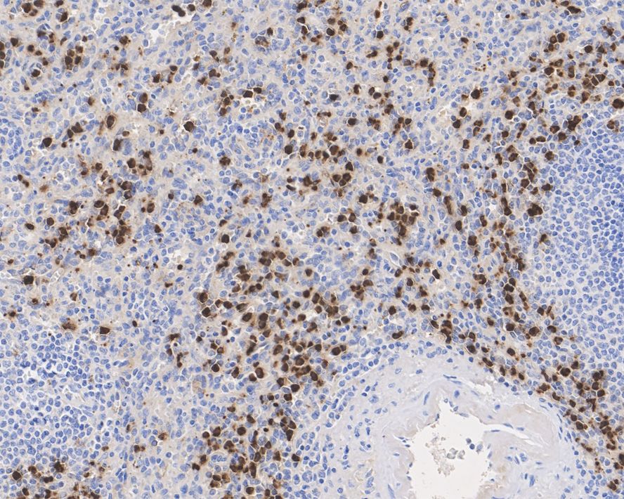
Immunohistochemical analysis of paraffin-embedded human spleen tissue with Rabbit anti-CLP antibody at 1/2,000 dilution. The section was pre-treated using heat mediated antigen retrieval with Tris-EDTA buffer (pH 9.0) for 20 minutes. The tissues were blocked in 1% BSA for 20 minutes at room temperature, washed with ddH2O and PBS, and then probed with the primary antibody at 1/2,000 dilution for 1 hour at room temperature. The detection was performed using an HRP conjugated compact polymer system. DAB was used as the chromogen. Tissues were counterstained with hematoxylin and mounted with DPX.IHC
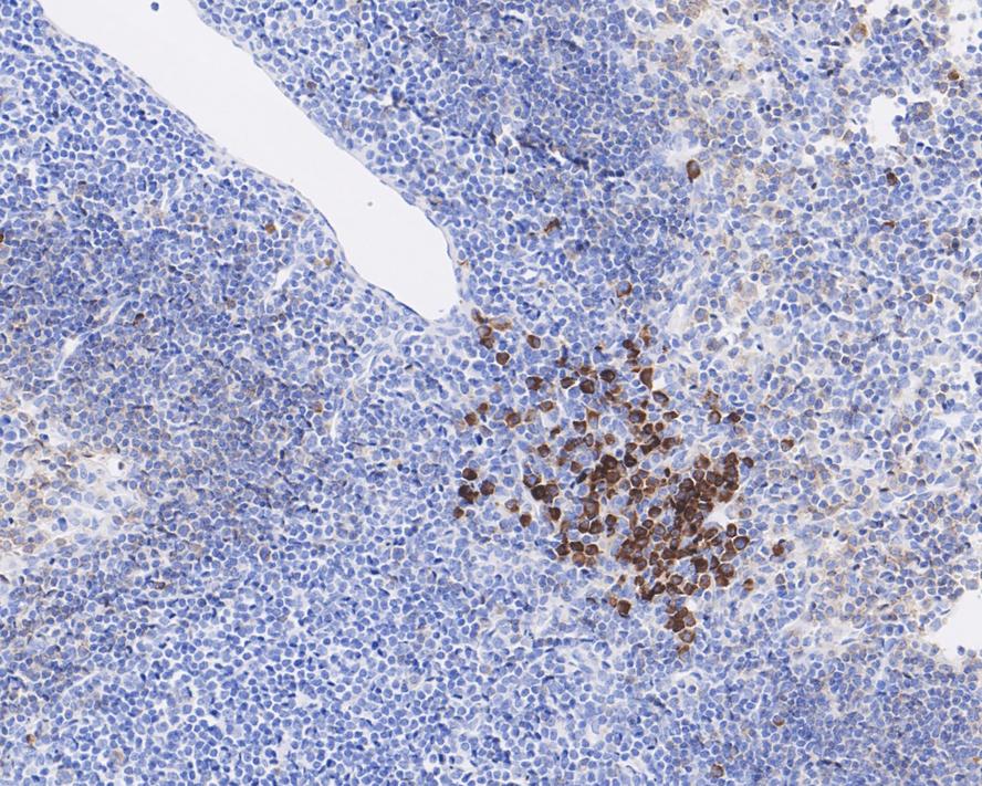
Immunohistochemical analysis of paraffin-embedded mouse spleen tissue with Rabbit anti-CLP antibody at 1/1,000 dilution. The section was pre-treated using heat mediated antigen retrieval with Tris-EDTA buffer (pH 9.0) for 20 minutes. The tissues were blocked in 1% BSA for 20 minutes at room temperature, washed with ddH2O and PBS, and then probed with the primary antibody at 1/1,000 dilution for 1 hour at room temperature. The detection was performed using an HRP conjugated compact polymer system. DAB was used as the chromogen. Tissues were counterstained with hematoxylin and mounted with DPX.IHC
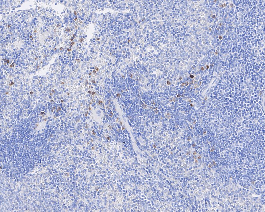
Immunohistochemical analysis of paraffin-embedded rat spleen tissue with Rabbit anti-CLP antibody at 1/1,000 dilution. The section was pre-treated using heat mediated antigen retrieval with Tris-EDTA buffer (pH 9.0) for 20 minutes. The tissues were blocked in 1% BSA for 20 minutes at room temperature, washed with ddH2O and PBS, and then probed with the primary antibody at 1/1,000 dilution for 1 hour at room temperature. The detection was performed using an HRP conjugated compact polymer system. DAB was used as the chromogen. Tissues were counterstained with hematoxylin and mounted with DPX.IF-P
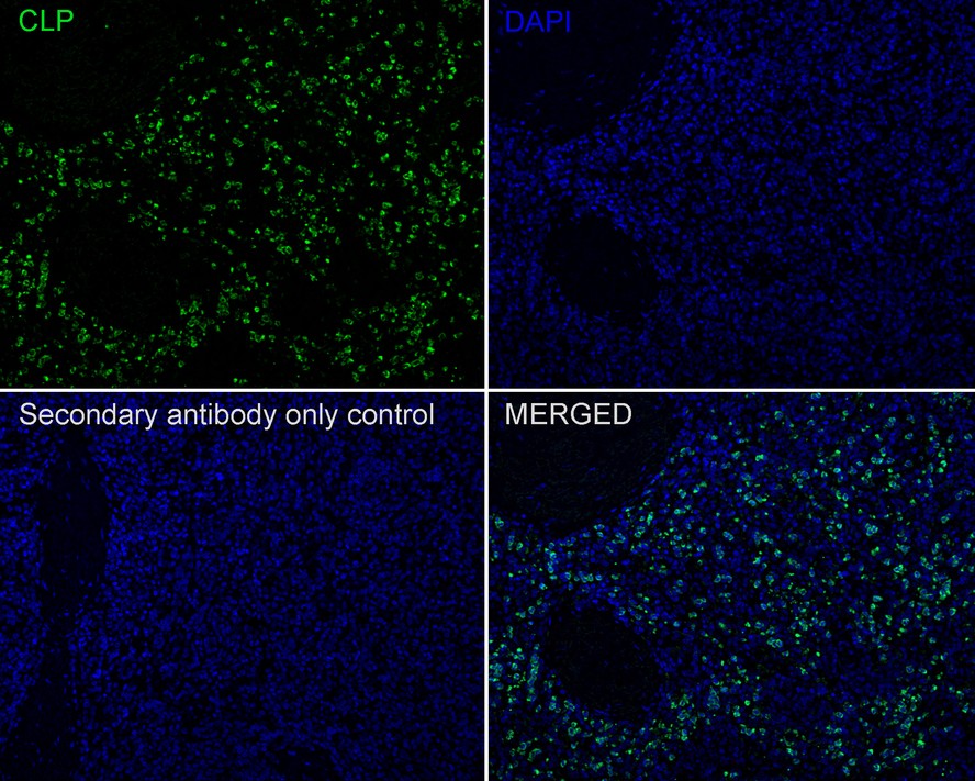
Immunofluorescence analysis of paraffin-embedded human spleen tissue labeling CLP with Rabbit anti-CLP antibody at 1/50 dilution. The section was pre-treated using heat mediated antigen retrieval with Tris-EDTA buffer (pH 9.0) for 20 minutes. The tissues were blocked in 10% negative goat serum for 1 hour at room temperature, washed with PBS, and then probed with the primary antibody (green) at 1/50 dilution overnight at 4 ℃, washed with PBS. Goat Anti-Rabbit IgG H&L (488) was used as the secondary antibody at 1/1,000 dilution. Nuclei were counterstained with DAPI (blue).| Product Name | CLP Recombinant Rabbit Monoclonal Antibody |
|---|---|
| Antibody Type | Primary Antibodies |
| Immunogen | Recombinant protein within human CLP aa 1-170 / 170. |
| Clonality | monoclonal |
|---|---|
| Isotype | IgG |
| Host Species | Rabbit |
| Tested Applications | IF-PIHC |
| IHC:1:1000-1:2000 IF-P:1:50 |
|
| Species Reactivity | HumanMouseRat |
| Concentration | 1mg/ml |
| Purification | Protein A |
| Gene Symbol | CAMP |
|---|---|
| Gene Synonyms | LL37 CAP18 CRAMP HSD26 CAP-18 FALL39 FALL-39 |
| Gene Full Name | cathelicidin antimicrobial peptide |
| Gene Summary | This gene encodes a member of an antimicrobial peptide family, characterized by a highly conserved N-terminal signal peptide containing a cathelin domain and a structurally variable cationic antimicrobial peptide, which is produced by extracellular proteolysis from the C-terminus. The protein plays an important role in innate immunity defense against viruses. In addition to its antibacterial, antifungal, and antiviral activities, the encoded protein functions in cell chemotaxis, immune mediator induction, and inflammatory response regulation. [provided by RefSeq, Sep 2021] |
| Alternative Names | 18 kDa cationic antimicrobial protein antibody Antibacterial peptide LL-37 antibody Antibacterial protein FALL-39 antibody CAMP antibody CAMP_HUMAN antibody CAP 18 antibody CAP-18 antibody CAP18 antibody Cathelicidin antimicrobial peptide antibody Cathelin-like protein antibody 18 kDa cationic antimicrobial protein antibody Antibacterial peptide LL-37 antibody Antibacterial protein FALL-39 antibody CAMP antibody CAMP_HUMAN antibody CAP 18 antibody CAP-18 antibody CAP18 antibody Cathelicidin antimicrobial peptide antibody Cathelin-like protein antibody Cathelin-related antimicrobial peptide antibody CATHL3 antibody Cationic antimicrobial protein 18-KD antibody CLP antibody Cnlp antibody Cramp antibody CRAMP mouse homolog of antibody FALL 39 antibody FALL-39 peptide antibiotic antibody FALL39 antibody hCAP 18 antibody hCAP-18 antibody hCAP18 antibody HSD26 antibody LL37 antibody MCLP antibody Peptide antibiotic PR-39 porcine homolog of antibody |
| Molecular Weight(MW) | 19kDa |
| Cellular Localization | Secreted, Vesicle. |
IHC
Immunohistochemical analysis of paraffin-embedded human spleen tissue with Rabbit anti-CLP antibody at 1/2,000 dilution. The section was pre-treated using heat mediated antigen retrieval with Tris-EDTA buffer (pH 9.0) for 20 minutes. The tissues were blocked in 1% BSA for 20 minutes at room temperature, washed with ddH2O and PBS, and then probed with the primary antibody at 1/2,000 dilution for 1 hour at room temperature. The detection was performed using an HRP conjugated compact polymer system. DAB was used as the chromogen. Tissues were counterstained with hematoxylin and mounted with DPX.
IHC
Immunohistochemical analysis of paraffin-embedded mouse spleen tissue with Rabbit anti-CLP antibody at 1/1,000 dilution. The section was pre-treated using heat mediated antigen retrieval with Tris-EDTA buffer (pH 9.0) for 20 minutes. The tissues were blocked in 1% BSA for 20 minutes at room temperature, washed with ddH2O and PBS, and then probed with the primary antibody at 1/1,000 dilution for 1 hour at room temperature. The detection was performed using an HRP conjugated compact polymer system. DAB was used as the chromogen. Tissues were counterstained with hematoxylin and mounted with DPX.
IHC
Immunohistochemical analysis of paraffin-embedded rat spleen tissue with Rabbit anti-CLP antibody at 1/1,000 dilution. The section was pre-treated using heat mediated antigen retrieval with Tris-EDTA buffer (pH 9.0) for 20 minutes. The tissues were blocked in 1% BSA for 20 minutes at room temperature, washed with ddH2O and PBS, and then probed with the primary antibody at 1/1,000 dilution for 1 hour at room temperature. The detection was performed using an HRP conjugated compact polymer system. DAB was used as the chromogen. Tissues were counterstained with hematoxylin and mounted with DPX.
IF-P
Immunofluorescence analysis of paraffin-embedded human spleen tissue labeling CLP with Rabbit anti-CLP antibody at 1/50 dilution. The section was pre-treated using heat mediated antigen retrieval with Tris-EDTA buffer (pH 9.0) for 20 minutes. The tissues were blocked in 10% negative goat serum for 1 hour at room temperature, washed with PBS, and then probed with the primary antibody (green) at 1/50 dilution overnight at 4 ℃, washed with PBS. Goat Anti-Rabbit IgG H&L (488) was used as the secondary antibody at 1/1,000 dilution. Nuclei were counterstained with DAPI (blue).| Application Notes | IHC:1:1000-1:2000 IF-P:1:50 |
|---|
| Form | Liquid |
|---|---|
| Storage Instructions | Store at +4℃ after thawing. Aliquot store at -20℃. Avoid repeated freeze / thaw cycles. |
| Storage Buffer | PBS (pH7.4), 0.1% BSA, 40% Glycerol. Preservative: 0.05% Sodium Azide. |


