WB
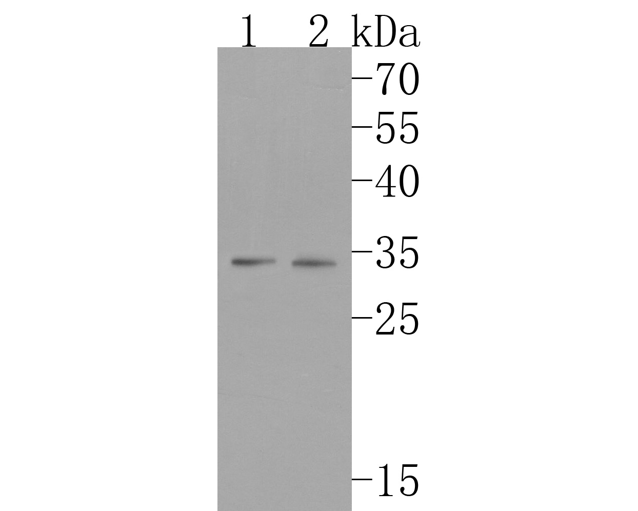
Western blot analysis of FHL1 on different lysates. Proteins were transferred to a PVDF membrane and blocked with 5% NFDM/TBST for 1 hour at room temperature. The primary antibody (1/500) was used in 5% NFDM/TBST at room temperature for 2 hours. Goat Anti-Rabbit IgG - HRP Secondary Antibody at 1:200,000 dilution was used for 1 hour at room temperature. Positive control: Lane 1: Jurkat cell lysate, Lane 2: Rat testis tissue lysate.IHC
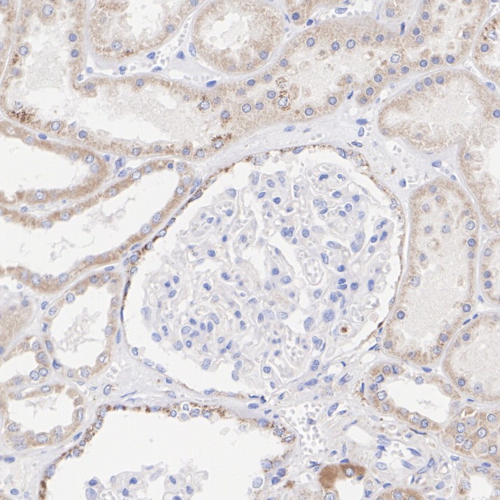
Immunohistochemical analysis of paraffin-embedded human kidney tissue with Rabbit anti-FHL1 antibody at 1/200 dilution. The section was pre-treated using heat mediated antigen retrieval with Tris-EDTA buffer (pH 9.0) for 20 minutes. The tissues were blocked in 1% BSA for 20 minutes at room temperature, washed with ddH2O and PBS, and then probed with the primary antibody at 1/200 dilution for 1 hour at room temperature. The detection was performed using an HRP conjugated compact polymer system. DAB was used as the chromogen. Tissues were counterstained with hematoxylin and mounted with DPX.IHC
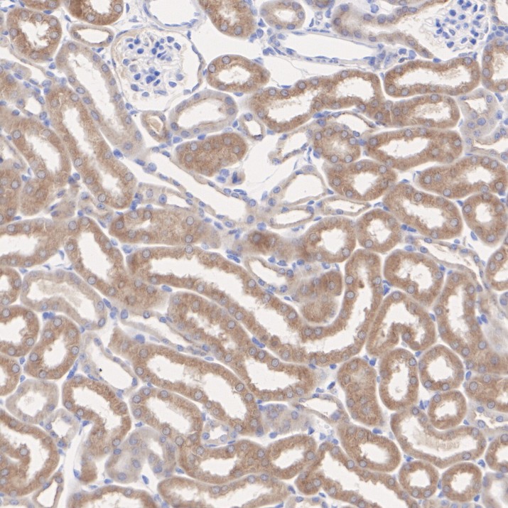
Immunohistochemical analysis of paraffin-embedded mouse kidney tissue with Rabbit anti-FHL1 antibody at 1/200 dilution. The section was pre-treated using heat mediated antigen retrieval with Tris-EDTA buffer (pH 9.0) for 20 minutes. The tissues were blocked in 1% BSA for 20 minutes at room temperature, washed with ddH2O and PBS, and then probed with the primary antibody at 1/200 dilution for 1 hour at room temperature. The detection was performed using an HRP conjugated compact polymer system. DAB was used as the chromogen. Tissues were counterstained with hematoxylin and mounted with DPX.IHC
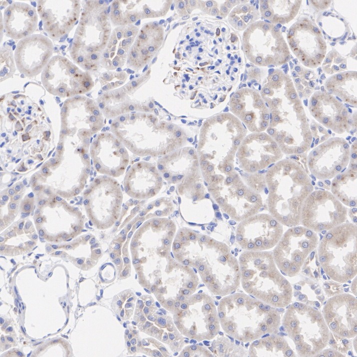
Immunohistochemical analysis of paraffin-embedded rat kidney tissue with Rabbit anti-FHL1 antibody at 1/200 dilution. The section was pre-treated using heat mediated antigen retrieval with Tris-EDTA buffer (pH 9.0) for 20 minutes. The tissues were blocked in 1% BSA for 20 minutes at room temperature, washed with ddH2O and PBS, and then probed with the primary antibody at 1/200 dilution for 1 hour at room temperature. The detection was performed using an HRP conjugated compact polymer system. DAB was used as the chromogen. Tissues were counterstained with hematoxylin and mounted with DPX.| Product Name | FHL1 Recombinant Rabbit Monoclonal Antibody |
|---|---|
| Antibody Type | Primary Antibodies |
| Immunogen | Synthetic peptide within human FHL1 aa 6-55/323. |
| Clonality | Monoclonal |
|---|---|
| Isotype | IgG |
| Host Species | Rabbit |
| Tested Applications | IHCWB |
| WB:1:500-1:1,000 IHC:1:200 |
|
| Species Reactivity | HumanMouseRat |
| Concentration | 1mg/ml |
| Purification | Protein A |
| Gene Symbol | FHL1 |
|---|---|
| Gene Synonyms | KYOT SLIM FCMSU FHL-1 FHL1A FHL1B FLH1A SLIM1 XMPMA RBMX1A RBMX1B SLIM-1 SLIMMER |
| Gene Full Name | This gene encodes a member of the four-and-a-half-LIM-only protein family. Family members contain two highly conserved, tandemly arranged, zinc finger domains with four highly conserved cysteines binding a zinc atom in each zinc finger. Expression of these family members occurs in a cell- and tissue-specific mode and these proteins are involved in many cellular processes. Mutations in this gene have been found in patients with Emery-Dreifuss muscular dystrophy. Multiple alternately spliced transcript variants which encode different protein isoforms have been described.[provided by RefSeq, Nov 2009] |
| Alternative Names | bA535K18.1 antibody FHL 1 antibody FHL 1B antibody FHL-1 antibody FHL1 antibody FHL1 protein antibody FHL1_HUMAN antibody FHL1A antibody FHL1B antibody FLH1A antibody bA535K18.1 antibody FHL 1 antibody FHL 1B antibody FHL-1 antibody FHL1 antibody FHL1 protein antibody FHL1_HUMAN antibody FHL1A antibody FHL1B antibody FLH1A antibody Four and a half LIM domains 1 antibody Four and a half LIM domains protein 1 antibody Four and a half Lin11 Isl 1 and Mec 3 domains 1 antibody four-and-a-half Lin11 antibody Isl-1 and Mec-3 antibody KYO T antibody KYOT mouse homolog of antibody LIM protein SLIMMER antibody MGC111107 antibody RAM14-1 antibody RBMX1A antibody RBMX1B antibody RBP associated molecule 14-1 antibody RP11-535K18.1 antibody Skeletal muscle LIM protein 1 antibody Skeletal muscle LIM-protein 1 antibody SLIM 1 antibody SLIM antibody SLIM-1 antibody SLIM1 antibody SLIMMER antibody XMPMA antibody |
| Molecular Weight(MW) | 36 kDa(Observed band size: 34 kDa) |
| Cellular Localization | Cytoplasm, Nucleus. |
WB
Western blot analysis of FHL1 on different lysates. Proteins were transferred to a PVDF membrane and blocked with 5% NFDM/TBST for 1 hour at room temperature. The primary antibody (1/500) was used in 5% NFDM/TBST at room temperature for 2 hours. Goat Anti-Rabbit IgG - HRP Secondary Antibody at 1:200,000 dilution was used for 1 hour at room temperature. Positive control: Lane 1: Jurkat cell lysate, Lane 2: Rat testis tissue lysate.
IHC
Immunohistochemical analysis of paraffin-embedded human kidney tissue with Rabbit anti-FHL1 antibody at 1/200 dilution. The section was pre-treated using heat mediated antigen retrieval with Tris-EDTA buffer (pH 9.0) for 20 minutes. The tissues were blocked in 1% BSA for 20 minutes at room temperature, washed with ddH2O and PBS, and then probed with the primary antibody at 1/200 dilution for 1 hour at room temperature. The detection was performed using an HRP conjugated compact polymer system. DAB was used as the chromogen. Tissues were counterstained with hematoxylin and mounted with DPX.
IHC
Immunohistochemical analysis of paraffin-embedded mouse kidney tissue with Rabbit anti-FHL1 antibody at 1/200 dilution. The section was pre-treated using heat mediated antigen retrieval with Tris-EDTA buffer (pH 9.0) for 20 minutes. The tissues were blocked in 1% BSA for 20 minutes at room temperature, washed with ddH2O and PBS, and then probed with the primary antibody at 1/200 dilution for 1 hour at room temperature. The detection was performed using an HRP conjugated compact polymer system. DAB was used as the chromogen. Tissues were counterstained with hematoxylin and mounted with DPX.
IHC
Immunohistochemical analysis of paraffin-embedded rat kidney tissue with Rabbit anti-FHL1 antibody at 1/200 dilution. The section was pre-treated using heat mediated antigen retrieval with Tris-EDTA buffer (pH 9.0) for 20 minutes. The tissues were blocked in 1% BSA for 20 minutes at room temperature, washed with ddH2O and PBS, and then probed with the primary antibody at 1/200 dilution for 1 hour at room temperature. The detection was performed using an HRP conjugated compact polymer system. DAB was used as the chromogen. Tissues were counterstained with hematoxylin and mounted with DPX.| Application Notes | WB:1:500-1:1,000 IHC:1:200 |
|---|
| Form | Liquid |
|---|---|
| Storage Instructions | Store at +4℃ after thawing. Aliquot store at -20℃. Avoid repeated freeze / thaw cycles. |
| Storage Buffer | 1*TBS (pH7.4), 0.05% BSA, 40% Glycerol. Preservative: 0.05% Sodium Azide. |


