WB
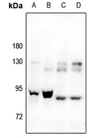
Western blot analysis of CAPRI expression in NIH3T3 (A), PC12 (B), HEK293T (C), SGC7901 (D) whole cell lysates.IHC
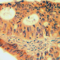
Immunohistochemical analysis of CAPRI staining in human lung cancer formalin fixed paraffin embedded tissue section. The section was pre-treated using heat mediated antigen retrieval with sodium citrate buffer (pH 6.0). The section was then incubated with the antibody at room temperature and detected using an HRP conjugated compact polymer system. DAB was used as the chromogen. The section was then counterstained with haematoxylin and mounted with DPX.ICC/IF
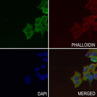
Immunofluorescent analysis of CAPRI staining in MCF7 cells. Formalin-fixed cells were permeabilized with 0.1% Triton X-100 in TBS for 5-10 minutes and blocked with 3% BSA-PBS for 30 minutes at room temperature. Cells were probed with the primary antibody in 3% BSA-PBS and incubated overnight at 4 °C in a hidified chamber. Cells were washed with PBST and incubated with a AF488-conjugated secondary antibody (green) in PBS at room temperature in the dark. Phalloidin - AF594 was used to stain Actin filaments (red). DAPI was used to stain the cell nuclei (blue).| Product Name | Anti-CAPRI Antibody |
|---|---|
| Antibody Type | Primary Antibodies |
| Immunogen | KLH-conjugated synthetic peptide encompassing a sequence within the center region of human CAPRI. The exact sequence is proprietary. |
| Clonality | Polyclonal |
|---|---|
| Isotype | IgG |
| Host Species | Rabbit |
| Tested Applications | ICC/IFIHCWB |
| WB:1:500-1:1000 IHC:1:50-1:100 ICC/IF:1:50-1:200 |
|
| Species Reactivity | HumanMouseRat |
| Concentration | 1mg/ml |
| Purification | Affinity purified |
| Gene Symbol | RASA4 |
|---|---|
| Gene Synonyms | GAPL CAPRI |
| Gene Full Name | RAS p21 protein activator 4 |
| Gene Summary | This gene encodes a member of the GAP1 family of GTPase-activating proteins that suppresses the Ras/mitogen-activated protein kinase pathway in response to Ca(2+). Stimuli that increase intracellular Ca(2+) levels result in the translocation of this protein to the plasma membrane, where it activates Ras GTPase activity. Consequently, Ras is converted from the active GTP-bound state to the inactive GDP-bound state and no longer activates downstream pathways that regulate gene expression, cell growth, and differentiation. Multiple transcript variants encoding different isoforms have been found for this gene. [provided by RefSeq, Jul 2008] |
| Alternative Names | CAPRI GAPL KIAA0538 Ras GTPase-activating protein 4 Calcium-promoted Ras inactivator Ras p21 protein activator 4 RasGAP-activating-like protein 2 |
| Molecular Weight(MW) | 90 kDa |
| Cellular Localization | Cell membrane,Cytoplasm,Membrane. |
WB
Western blot analysis of CAPRI expression in NIH3T3 (A), PC12 (B), HEK293T (C), SGC7901 (D) whole cell lysates.
IHC
Immunohistochemical analysis of CAPRI staining in human lung cancer formalin fixed paraffin embedded tissue section. The section was pre-treated using heat mediated antigen retrieval with sodium citrate buffer (pH 6.0). The section was then incubated with the antibody at room temperature and detected using an HRP conjugated compact polymer system. DAB was used as the chromogen. The section was then counterstained with haematoxylin and mounted with DPX.
ICC/IF
Immunofluorescent analysis of CAPRI staining in MCF7 cells. Formalin-fixed cells were permeabilized with 0.1% Triton X-100 in TBS for 5-10 minutes and blocked with 3% BSA-PBS for 30 minutes at room temperature. Cells were probed with the primary antibody in 3% BSA-PBS and incubated overnight at 4 °C in a hidified chamber. Cells were washed with PBST and incubated with a AF488-conjugated secondary antibody (green) in PBS at room temperature in the dark. Phalloidin - AF594 was used to stain Actin filaments (red). DAPI was used to stain the cell nuclei (blue).| Application Notes | WB:1:500-1:1000 IHC:1:50-1:100 ICC/IF:1:50-1:200 |
|---|
| Form | Liquid |
|---|---|
| Storage Instructions | Shipped at 4°C. Upon delivery aliquot and store at -20°C for one year. Avoid freeze/thaw cycles. |
| Storage Buffer | Liquid in 0.42% Potassium phosphate, 0.87% Sodium chloride, pH 7.3, 30% glycerol, and 0.01% sodium azide. |


