WB
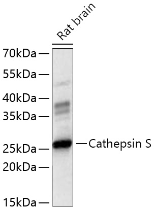
Western blot analysis of lysates from Rat brain using Cathepsin S Rabbit pAb at 1:3000 dilution incubated at room temperature for 1.5 hours. Secondary antibody: HRP-conjugated Goat anti-Rabbit IgG (H+L) at 1:10000 dilution. Lysates/proteins: 25 μg per lane. Blocking buffer: 3% nonfat dry milk in TBST. Detection: ECL Basic Kit. Exposure time: 60s.IHC
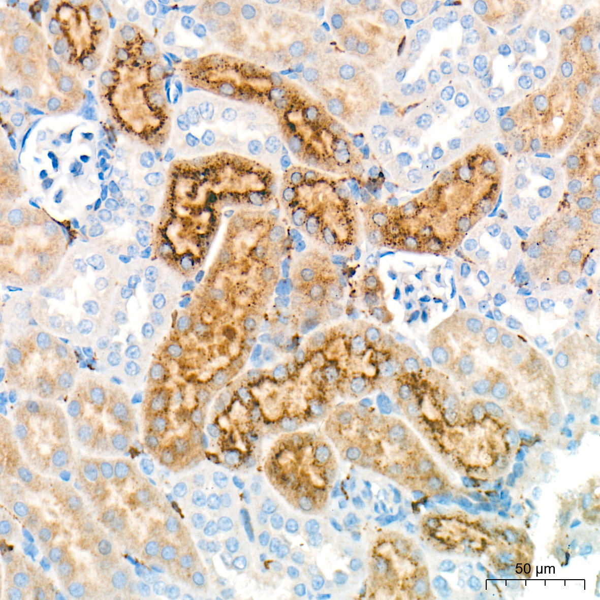
Immunohistochemistry analysis of paraffin embedded Mouse kidney tissue using Cathepsin S Rabbit pAb at a dilution of 1:400 (40x lens). High pressure antigen retrieval performed with 0.01M Tris EDTA Buffer (pH 9.0) prior to IHC staining.IHC
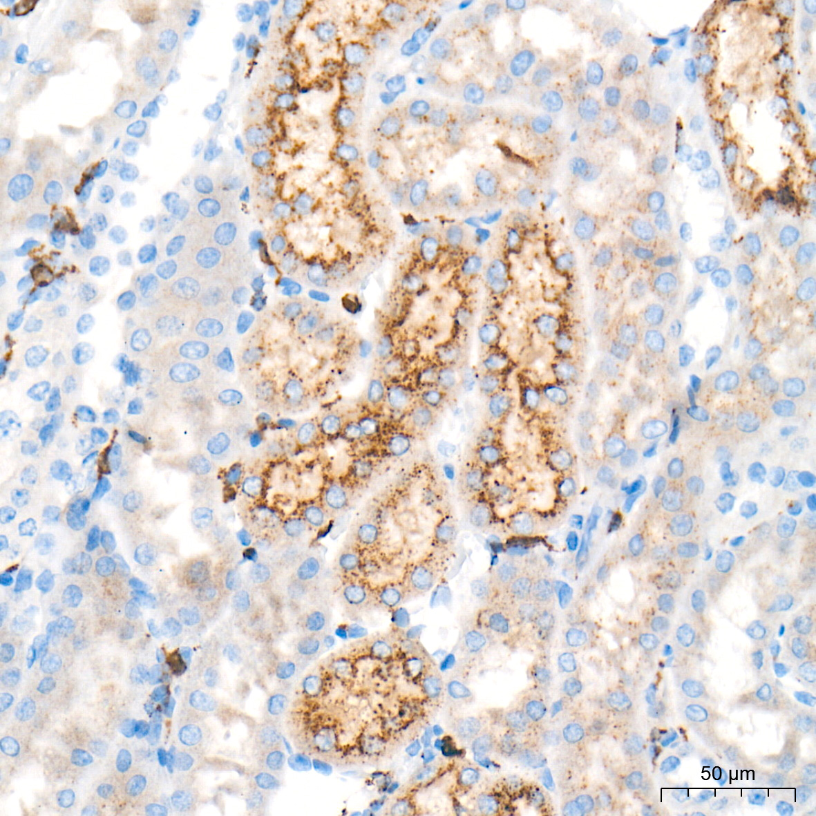
Immunohistochemistry analysis of paraffin embedded Rat kidney tissue using Cathepsin S Rabbit pAb at a dilution of 1:400 (40x lens). High pressure antigen retrieval performed with 0.01M Tris-EDTA Buffer (pH 9.0) prior to IHC staining.ICC/IF
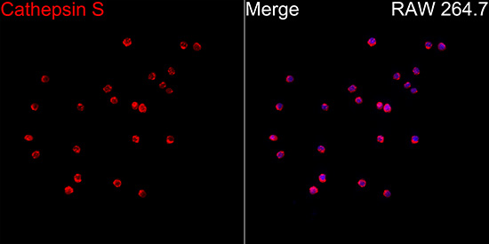
Immunofluorescence analysis of RAW 264.7 cells using Cathepsin S Rabbit pAb at a dilution of 1:200 (40x lens). Secondary antibody: Cy3-conjugated Goat anti-Rabbit IgG (H+L) at 1:500 dilution. Blue: DAPI for nuclear staining.IF-P
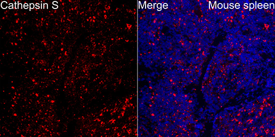
Immunofluorescence analysis of Mouse spleen tissue using Cathepsin S Rabbit pAb at a dilution of 1:200 (40x lens). Secondary antibody: Cy3-conjugated Goat anti-Rabbit IgG (H+L)at 1:500 dilution. Blue: DAPI for nuclear staining. High pressure antigen retrieval performed with 0.01M Citrate Buffer (pH 6.0) prior to IF staining.IF-P
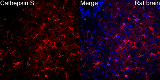
Immunofluorescence analysis of Rat brain tissue using Cathepsin S Rabbit pAb at a dilution of 1:200 (40x lens). Secondary antibody: Cy3-conjugated Goat anti-Rabbit IgG (H+L)at 1:500 dilution. Blue: DAPI for nuclear staining. High pressure antigen retrieval performed with 0.01M Citrate Buffer (pH 6.0) prior to IF staining.| Product Name | Cathepsin S Rabbit pAb |
|---|---|
| Antibody Type | Primary Antibodies |
| Immunogen | Recombinant protein. |
| Clonality | Polyclonal |
|---|---|
| Isotype | IgG |
| Host Species | Rabbit |
| Tested Applications | ICC/IFIF-PIHCWB |
| WB:1:1000-1:3000 IHC:1:200-1:800 ICC/IF:1:100-1:400 IF-P:1:100-1:400 |
|
| Species Reactivity | MouseRat |
| Concentration | 1mg/ml |
| Purification | Affinity purified |
| Gene Symbol | Ctss |
|---|---|
| Gene Synonyms | Cats |
| Gene Full Name | cathepsin S |
| Gene Summary | This gene encodes a member of the peptidase C1 (papain) family of cysteine proteases. Alternative splicing results in multiple transcript variants, which encode preproproteins that are proteolytically processed to generate mature protein products. This enzyme is secreted by antigen-presenting cells during inflammation and may induce pain and itch via activation of G-protein coupled receptors. Homozygous knockout mice for this gene exhibit impaired wound healing, reduced tumorigenesis in a pancreatic cancer model, and reduced pathogenesis in a myasthenia gravis model. [provided by RefSeq, Aug 2015] |
| Alternative Names | Cats |
| Molecular Weight(MW) | 38kDa(Observed MW 25kDa) |
| Cellular Localization | Lysosome, Secreted, Cytoplasmic vesicle, phagosome. |
WB
Western blot analysis of lysates from Rat brain using Cathepsin S Rabbit pAb at 1:3000 dilution incubated at room temperature for 1.5 hours. Secondary antibody: HRP-conjugated Goat anti-Rabbit IgG (H+L) at 1:10000 dilution. Lysates/proteins: 25 μg per lane. Blocking buffer: 3% nonfat dry milk in TBST. Detection: ECL Basic Kit. Exposure time: 60s.
IHC
Immunohistochemistry analysis of paraffin embedded Mouse kidney tissue using Cathepsin S Rabbit pAb at a dilution of 1:400 (40x lens). High pressure antigen retrieval performed with 0.01M Tris EDTA Buffer (pH 9.0) prior to IHC staining.
IHC
Immunohistochemistry analysis of paraffin embedded Rat kidney tissue using Cathepsin S Rabbit pAb at a dilution of 1:400 (40x lens). High pressure antigen retrieval performed with 0.01M Tris-EDTA Buffer (pH 9.0) prior to IHC staining.
ICC/IF
Immunofluorescence analysis of RAW 264.7 cells using Cathepsin S Rabbit pAb at a dilution of 1:200 (40x lens). Secondary antibody: Cy3-conjugated Goat anti-Rabbit IgG (H+L) at 1:500 dilution. Blue: DAPI for nuclear staining.
IF-P
Immunofluorescence analysis of Mouse spleen tissue using Cathepsin S Rabbit pAb at a dilution of 1:200 (40x lens). Secondary antibody: Cy3-conjugated Goat anti-Rabbit IgG (H+L)at 1:500 dilution. Blue: DAPI for nuclear staining. High pressure antigen retrieval performed with 0.01M Citrate Buffer (pH 6.0) prior to IF staining.
IF-P
Immunofluorescence analysis of Rat brain tissue using Cathepsin S Rabbit pAb at a dilution of 1:200 (40x lens). Secondary antibody: Cy3-conjugated Goat anti-Rabbit IgG (H+L)at 1:500 dilution. Blue: DAPI for nuclear staining. High pressure antigen retrieval performed with 0.01M Citrate Buffer (pH 6.0) prior to IF staining.| Application Notes | WB:1:1000-1:3000 IHC:1:200-1:800 ICC/IF:1:100-1:400 IF-P:1:100-1:400 |
|---|
| Form | Liquid |
|---|---|
| Storage Instructions | Store at -20℃. Avoid freeze / thaw cycles. |
| Storage Buffer | Buffer: PBS with 0.05% proclin300, 0.05% BSA, 50% glycerol, pH7.3. |


