WB
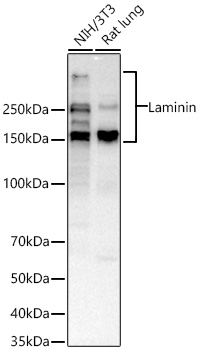
Western blot analysis of various lysates using Laminin Rabbit pAb at 1:1000 dilution incubated overnight at 4℃. Secondary antibody: HRP-conjugated Goat anti-Rabbit IgG (H+L) at 1:10000 dilution. Lysates/proteins: 25 μg per lane. Blocking buffer: 3% nonfat dry milk in TBST. Detection: ECL Basic Kit. Exposure time: 45s.IHC
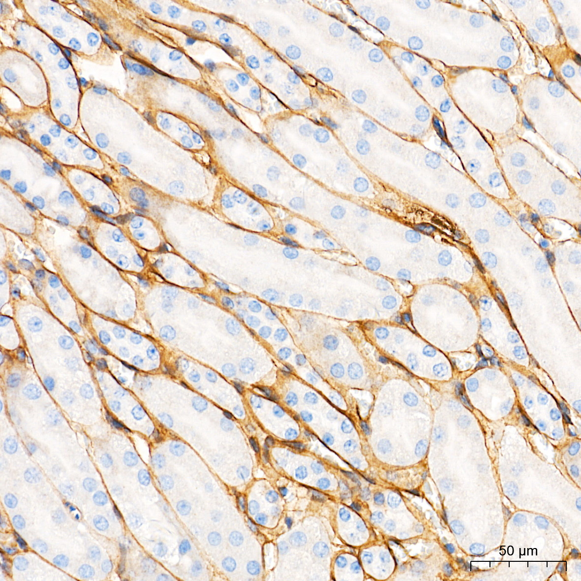
Immunohistochemistry analysis of paraffin embedded Mouse kidney tissue using Laminin Rabbit pAb at a dilution of 1:200 (40x lens). High pressure antigen retrieval performed with 0.01M Tris-EDTA Buffer (pH 9.0) prior to IHC staining.IHC
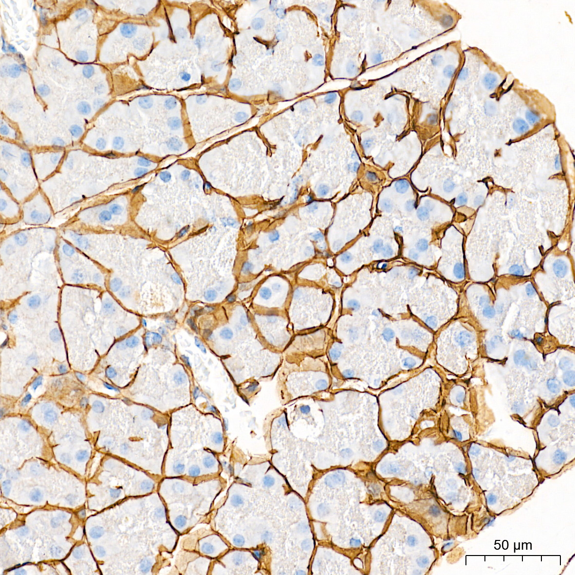
Immunohistochemistry analysis of paraffin embedded Mouse pancreas tissue using Laminin Rabbit pAb at a dilution of 1:200 (40x lens). High pressure antigen retrieval performed with 0.01M Tris-EDTA Buffer (pH 9.0) prior to IHC staining.ICC/IF
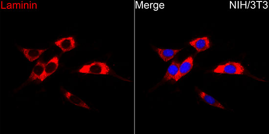
Immunofluorescence analysis of NIH/3T3 cells using Laminin Rabbit pAb at a dilution of 1:200 (40x lens). Secondary antibody: Cy3-conjugated Goat anti-Rabbit IgG (H+L)at 1:500 dilution. Blue: DAPI for nuclear staining.IF-P
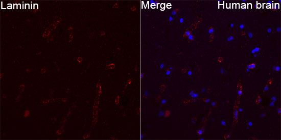
Immunofluorescence analysis of Human brain tissue using Laminin Rabbit pAb at a dilution of 1:200 (40x lens). Secondary antibody: Cy3-conjugated Goat anti-Rabbit IgG (H+L) at 1:500 dilution. Blue: DAPI for nuclear staining. High pressure antigen retrieval performed with 0.01M Citrate Buffer (pH 6.0) prior to IF staining.IF-P
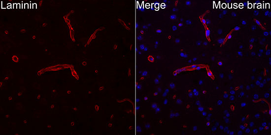
Immunofluorescence analysis of Mouse brain tissue using Laminin Rabbit pAb at a dilution of 1:200 (40x lens). Secondary antibody: Cy3-conjugated Goat anti-Rabbit IgG (H+L) at 1:500 dilution. Blue: DAPI for nuclear staining. High pressure antigen retrieval performed with 0.01M Citrate Buffer (pH 6.0) prior to IF staining.| Product Name | Laminin Rabbit pAb |
|---|---|
| Antibody Type | Primary Antibodies |
| Immunogen | Recombinant protein. |
| Clonality | Polyclonal |
|---|---|
| Isotype | IgG |
| Host Species | Rabbit |
| Tested Applications | ICC/IFIF-PIHCWB |
| WB:1:1000-1:2000 IHC:1:200-1:800 ICC/IF:1:100-1:400 IF-P:1:100-1:400 |
|
| Species Reactivity | HumanMouseRat |
| Concentration | 1mg/ml |
| Purification | Affinity purified |
| Gene Symbol | LAMA2/LAMB2/LAMB1/LAMC1/LAMC3 |
|---|---|
| Gene Synonyms | ARP Arp1 ARL18 |
| Gene Summary | Laminin, an extracellular protein, is a major component of the basement membrane. It is thought to mediate the attachment, migration, and organization of cells into tissues during embryonic development by interacting with other extracellular matrix components. It is composed of three subunits, alpha, beta, and gamma, which are bound to each other by disulfide bonds into a cross-shaped molecule. This gene encodes the alpha 2 chain, which constitutes one of the subunits of laminin 2 (merosin) and laminin 4 (s-merosin). Mutations in this gene have been identified as the cause of congenital merosin-deficient muscular dystrophy. Two transcript variants encoding different proteins have been found for this gene. [provided by RefSeq, Jul 2008] |
| Alternative Names | ARP Arp1 ARL18 |
| Molecular Weight(MW) | 129-404kDa |
| Cellular Localization | Golgi apparatus, trans-Golgi network. |
WB
Western blot analysis of various lysates using Laminin Rabbit pAb at 1:1000 dilution incubated overnight at 4℃. Secondary antibody: HRP-conjugated Goat anti-Rabbit IgG (H+L) at 1:10000 dilution. Lysates/proteins: 25 μg per lane. Blocking buffer: 3% nonfat dry milk in TBST. Detection: ECL Basic Kit. Exposure time: 45s.
IHC
Immunohistochemistry analysis of paraffin embedded Mouse kidney tissue using Laminin Rabbit pAb at a dilution of 1:200 (40x lens). High pressure antigen retrieval performed with 0.01M Tris-EDTA Buffer (pH 9.0) prior to IHC staining.
IHC
Immunohistochemistry analysis of paraffin embedded Mouse pancreas tissue using Laminin Rabbit pAb at a dilution of 1:200 (40x lens). High pressure antigen retrieval performed with 0.01M Tris-EDTA Buffer (pH 9.0) prior to IHC staining.
ICC/IF
Immunofluorescence analysis of NIH/3T3 cells using Laminin Rabbit pAb at a dilution of 1:200 (40x lens). Secondary antibody: Cy3-conjugated Goat anti-Rabbit IgG (H+L)at 1:500 dilution. Blue: DAPI for nuclear staining.
IF-P
Immunofluorescence analysis of Human brain tissue using Laminin Rabbit pAb at a dilution of 1:200 (40x lens). Secondary antibody: Cy3-conjugated Goat anti-Rabbit IgG (H+L) at 1:500 dilution. Blue: DAPI for nuclear staining. High pressure antigen retrieval performed with 0.01M Citrate Buffer (pH 6.0) prior to IF staining.
IF-P
Immunofluorescence analysis of Mouse brain tissue using Laminin Rabbit pAb at a dilution of 1:200 (40x lens). Secondary antibody: Cy3-conjugated Goat anti-Rabbit IgG (H+L) at 1:500 dilution. Blue: DAPI for nuclear staining. High pressure antigen retrieval performed with 0.01M Citrate Buffer (pH 6.0) prior to IF staining.| Application Notes | WB:1:1000-1:2000 IHC:1:200-1:800 ICC/IF:1:100-1:400 IF-P:1:100-1:400 |
|---|
| Form | Liquid |
|---|---|
| Storage Instructions | Store at -20℃. Avoid freeze / thaw cycles. |
| Storage Buffer | Buffer: PBS with 0.05% proclin300, 0.05% BSA, 50% glycerol, pH7.3. |


