WB
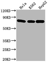
Positive WB detected in: Hela whole cell lysate, K562 whole cell lysate, HepG2 whole cell lysate, All lanes HSPA8 antibody at 1:2000, Secondary Goat polyclonal to mouse IgG at 1/50000 dilution, Exposure time: 5min.IHC
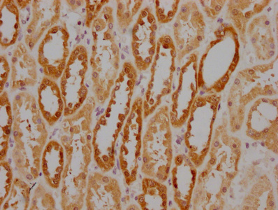
IHC image of HSPA8 Monoclonal Antibody diluted at 1:220 and staining in paraffin-embedded human kidney tissue performed on a Leica BondTM system. After dewaxing and hydration, antigen retrieval was mediated by high pressure in a citrate buffer (pH 6.0). Section was blocked with 10% normal goat serum 30min at RT. Then primary antibody (1% BSA) was incubated at 4°C overnight. The primary is detected by a biotinylated secondary antibody and visualized using an HRP conjugated SP system.IHC
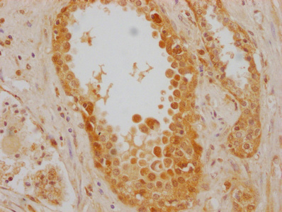
IHC image of HSPA8 Monoclonal Antibody diluted at 1:220 and staining in paraffin-embedded human prostate cancer performed on a Leica BondTM system. After dewaxing and hydration, antigen retrieval was mediated by high pressure in a citrate buffer (pH 6.0). Section was blocked with 10% normal goat serum 30min at RT. Then primary antibody (1% BSA) was incubated at 4°C overnight. The primary is detected by a biotinylated secondary antibody and visualized using an HRP conjugated SP system.ICC/IF
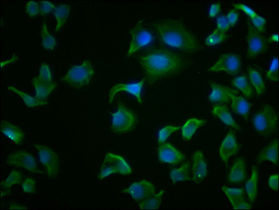
Immunofluorescence staining of Hela cells with HSPA8 Monoclonal Antibody at 1:100, counter-stained with DAPI. The cells were fixed in 4% formaldehyde and blocked in 10% normal Goat Serum. The cells were then incubated with the antibody overnight at 4°C. Nuclear DNA was labeled in blue with DAPI. The secondary antibody was FITC-conjugated AffiniPure Goat Anti-Mouse IgG (H+L).FC
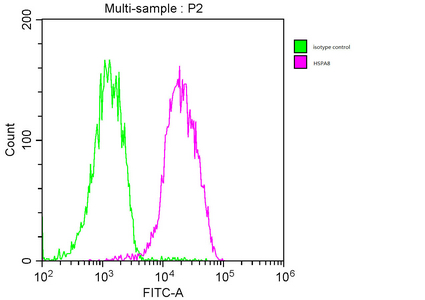
Overlay histogram showing MCF-7 cells stained with HSPA8 Monoclonal Antibody (red line). The cells were fixed with 70% Ethylalcohol (18h) and then incubated in 10% normal goat serum to block non-specific protein-protein interactions followed by the primary antibody at 1:200 for 1 h at 4°C. The secondary antibody used was FITC goat anti-mouse IgG(H+L) at 1/100 dilution for 30min at 4°C. Isotype control antibody (green line) was mouse IgG1 used under the same conditions. Acquisition of >10,000 events was performed.| Product Name | Mouse anti-HSPA8 Monoclonal antibody |
|---|---|
| Antibody Type | Primary Antibodies |
| Immunogen | Recombinant Human DPP9 protein |
| Clonality | Monoclonal |
|---|---|
| Isotype | IgG1 |
| Host Species | Mouse |
| Tested Applications | FCICC/IFIHCWB |
| WB:1:5000-1:30000 IHC:1:100-1:500 ICC/IF:1:100-1:500 FC:1:100-1:300 |
|
| Species Reactivity | Human |
| Concentration | 1mg/ml |
| Purification | Affinity purified |
| Gene Symbol | HSPA8 |
|---|---|
| Gene Synonyms | LAP1 HSC54 HSC70 HSC71 HSP71 HSP73 LAP-1 NIP71 HEL-33 HSPA10 HEL-S-72p |
| Gene Full Name | heat shock protein family A (Hsp70) member 8 |
| Gene Summary | This gene encodes a member of the heat shock protein 70 family, which contains both heat-inducible and constitutively expressed members. This protein belongs to the latter group, which are also referred to as heat-shock cognate proteins. It functions as a chaperone, and binds to nascent polypeptides to facilitate correct folding. It also functions as an ATPase in the disassembly of clathrin-coated vesicles during transport of membrane components through the cell. Alternatively spliced transcript variants encoding different isoforms have been found for this gene. [provided by RefSeq, Aug 2011] |
| Alternative Names | 2410008N15Rik antibody Constitutive heat shock protein 70 antibody Epididymis luminal protein 33 antibody Epididymis secretory sperm binding protein Li 72p antibody Heat shock 70 kDa protein 8 antibody Heat shock 70kD protein 10 antibody Heat shock 70kD protein 8 antibody Heat shock 70kDa protein 8 antibody Heat shock cognate 71 kDa protein antibody Heat shock cognate protein 54 antibody Heat shock cognate protein 71 kDa antibody Heat shock protein 8 antibody Heat shock protein A8 antibody Heat shock protein family A (Hsp70) member 8 antibody Heat-shock70-KD protein 10 formerly antibody HEL 33 antibody HEL S 72p antibody HSC54 antibody HSC71 antibody Hsc73 antibody HSP71 antibody HSP73 antibody HSP7C_HUMAN antibody HSPA10 antibody HSPA8 antibody LAP1 antibody Lipopolysaccharide associated protein 1 antibody LPS associated protein 1 antibody LPS associated protein antibody MGC102007 antibody MGC106514 antibody MGC114311 antibody MGC118485 antibody MGC131511 antibody MGC29929 antibody N-myristoyltransferase inhibitor protein 71 antibody NIP71 antibody |
| Molecular Weight(MW) | 71 kDa |
| Cellular Localization | Cytoplasm, Nucleus, Cell membrane, Melanosome. |
WB
Positive WB detected in: Hela whole cell lysate, K562 whole cell lysate, HepG2 whole cell lysate, All lanes HSPA8 antibody at 1:2000, Secondary Goat polyclonal to mouse IgG at 1/50000 dilution, Exposure time: 5min.
IHC
IHC image of HSPA8 Monoclonal Antibody diluted at 1:220 and staining in paraffin-embedded human kidney tissue performed on a Leica BondTM system. After dewaxing and hydration, antigen retrieval was mediated by high pressure in a citrate buffer (pH 6.0). Section was blocked with 10% normal goat serum 30min at RT. Then primary antibody (1% BSA) was incubated at 4°C overnight. The primary is detected by a biotinylated secondary antibody and visualized using an HRP conjugated SP system.
IHC
IHC image of HSPA8 Monoclonal Antibody diluted at 1:220 and staining in paraffin-embedded human prostate cancer performed on a Leica BondTM system. After dewaxing and hydration, antigen retrieval was mediated by high pressure in a citrate buffer (pH 6.0). Section was blocked with 10% normal goat serum 30min at RT. Then primary antibody (1% BSA) was incubated at 4°C overnight. The primary is detected by a biotinylated secondary antibody and visualized using an HRP conjugated SP system.
ICC/IF
Immunofluorescence staining of Hela cells with HSPA8 Monoclonal Antibody at 1:100, counter-stained with DAPI. The cells were fixed in 4% formaldehyde and blocked in 10% normal Goat Serum. The cells were then incubated with the antibody overnight at 4°C. Nuclear DNA was labeled in blue with DAPI. The secondary antibody was FITC-conjugated AffiniPure Goat Anti-Mouse IgG (H+L).
FC
Overlay histogram showing MCF-7 cells stained with HSPA8 Monoclonal Antibody (red line). The cells were fixed with 70% Ethylalcohol (18h) and then incubated in 10% normal goat serum to block non-specific protein-protein interactions followed by the primary antibody at 1:200 for 1 h at 4°C. The secondary antibody used was FITC goat anti-mouse IgG(H+L) at 1/100 dilution for 30min at 4°C. Isotype control antibody (green line) was mouse IgG1 used under the same conditions. Acquisition of >10,000 events was performed.| Application Notes | WB:1:5000-1:30000 IHC:1:100-1:500 ICC/IF:1:100-1:500 FC:1:100-1:300 |
|---|
| Form | Liquid |
|---|---|
| Storage Instructions | Upon receipt, store at -20°C or -80°C. Avoid repeated freeze. |
| Storage Buffer | 0.03% Proclin 300 Constituents: 50% Glycerol, 0.01M PBS, PH 7.4 |


