IHC
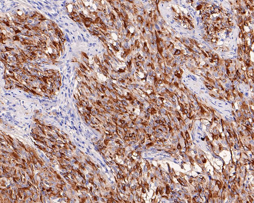
Immunohistochemical analysis of paraffin-embedded human medullary thyroid carcinoma tissue with Rabbit anti-Calcitonin antibody at 1/20,000 dilution. The section was pre-treated using heat mediated antigen retrieval with Tris-EDTA buffer (pH 9.0) for 20 minutes. The tissues were blocked in 1% BSA for 20 minutes at room temperature, washed with ddH2O and PBS, and then probed with the primary antibody at 1/20,000 dilution for 1 hour at room temperature. The detection was performed using an HRP conjugated compact polymer system. DAB was used as the chromogen. Tissues were counterstained with hematoxylin and mounted with DPX.IHC
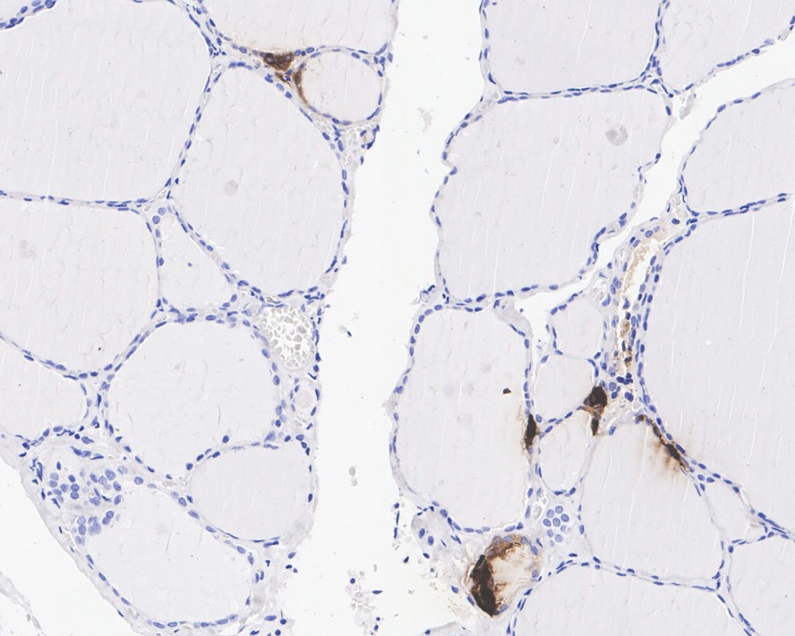
Immunohistochemical analysis of paraffin-embedded human thyroid tissue with Rabbit anti-Calcitonin antibody at 1/2,000 dilution. The section was pre-treated using heat mediated antigen retrieval with Tris-EDTA buffer (pH 9.0) for 20 minutes. The tissues were blocked in 1% BSA for 20 minutes at room temperature, washed with ddH2O and PBS, and then probed with the primary antibody at 1/2,000 dilution for 1 hour at room temperature. The detection was performed using an HRP conjugated compact polymer system. DAB was used as the chromogen. Tissues were counterstained with hematoxylin and mounted with DPX.IHC
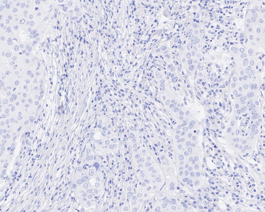
Immunohistochemical analysis of paraffin-embedded human breast cancer tissue (negative) with Rabbit anti-Calcitonin antibody at 1/2,000 dilution. The section was pre-treated using heat mediated antigen retrieval with Tris-EDTA buffer (pH 9.0) for 20 minutes. The tissues were blocked in 1% BSA for 20 minutes at room temperature, washed with ddH2O and PBS, and then probed with the primary antibody at 1/2,000 dilution for 1 hour at room temperature. The detection was performed using an HRP conjugated compact polymer system. DAB was used as the chromogen. Tissues were counterstained with hematoxylin and mounted with DPX.IHC
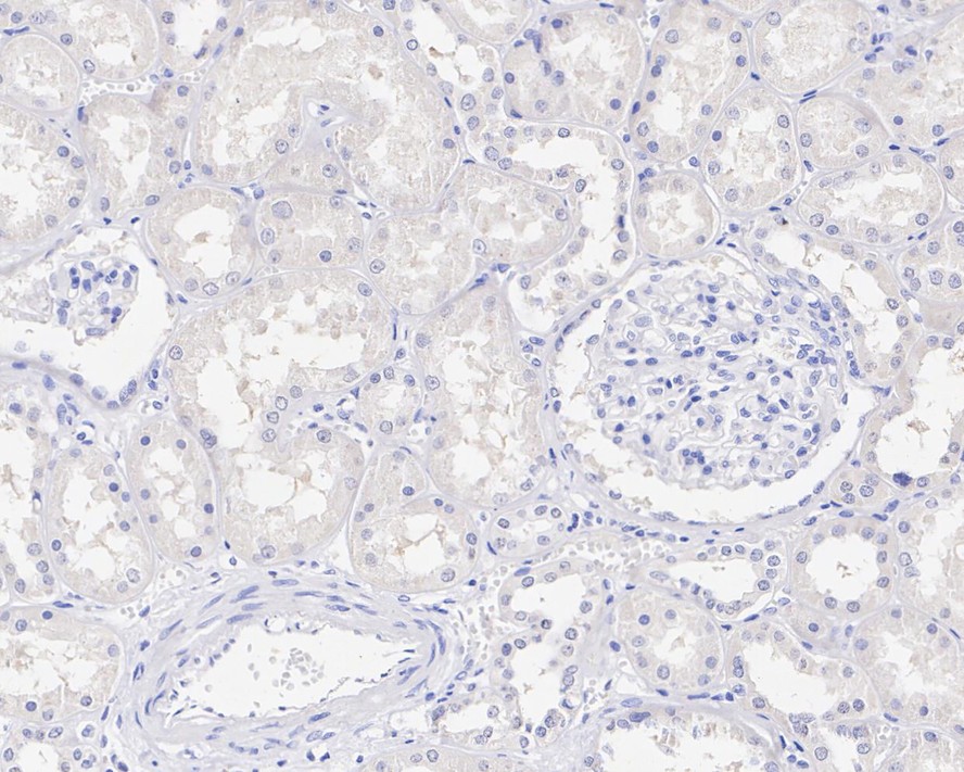
Immunohistochemical analysis of paraffin-embedded human kidney tissue (negative) with Rabbit anti-Calcitonin antibody at 1/2,000 dilution. The section was pre-treated using heat mediated antigen retrieval with Tris-EDTA buffer (pH 9.0) for 20 minutes. The tissues were blocked in 1% BSA for 20 minutes at room temperature, washed with ddH2O and PBS, and then probed with the primary antibody at 1/2,000 dilution for 1 hour at room temperature. The detection was performed using an HRP conjugated compact polymer system. DAB was used as the chromogen. Tissues were counterstained with hematoxylin and mounted with DPX.ICC/IF
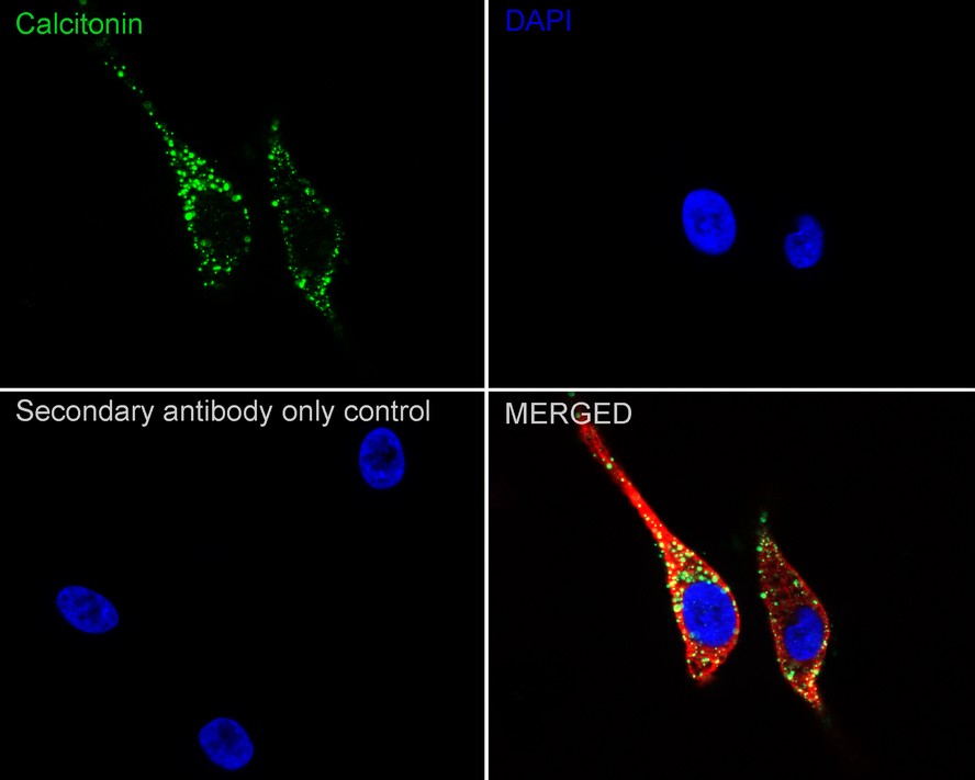
Immunocytochemistry analysis of TT cells labeling Calcitonin with Rabbit anti-Calcitonin antibody at 1/100 dilution. Cells were fixed in 4% paraformaldehyde for 20 minutes at room temperature, permeabilized with 0.1% Triton X-100 in PBS for 5 minutes at room temperature, then blocked with 1% BSA in 10% negative goat serum for 1 hour at room temperature. Cells were then incubated with Rabbit anti-Calcitonin antibody at 1/100 dilution in 1% BSA in PBST overnight at 4 ℃. Goat Anti-Rabbit IgG H&L (488) was used as the secondary antibody at 1/1,000 dilution. PBS instead of the primary antibody was used as the secondary antibody only control. Nuclear DNA was labelled in blue with DAPI. Beta tubulin (red) was stained at 1/100 dilution overnight at +4℃. Goat Anti-Mouse IgG H&L (594) was used as the secondary antibody at 1/1,000 dilution.IF-P
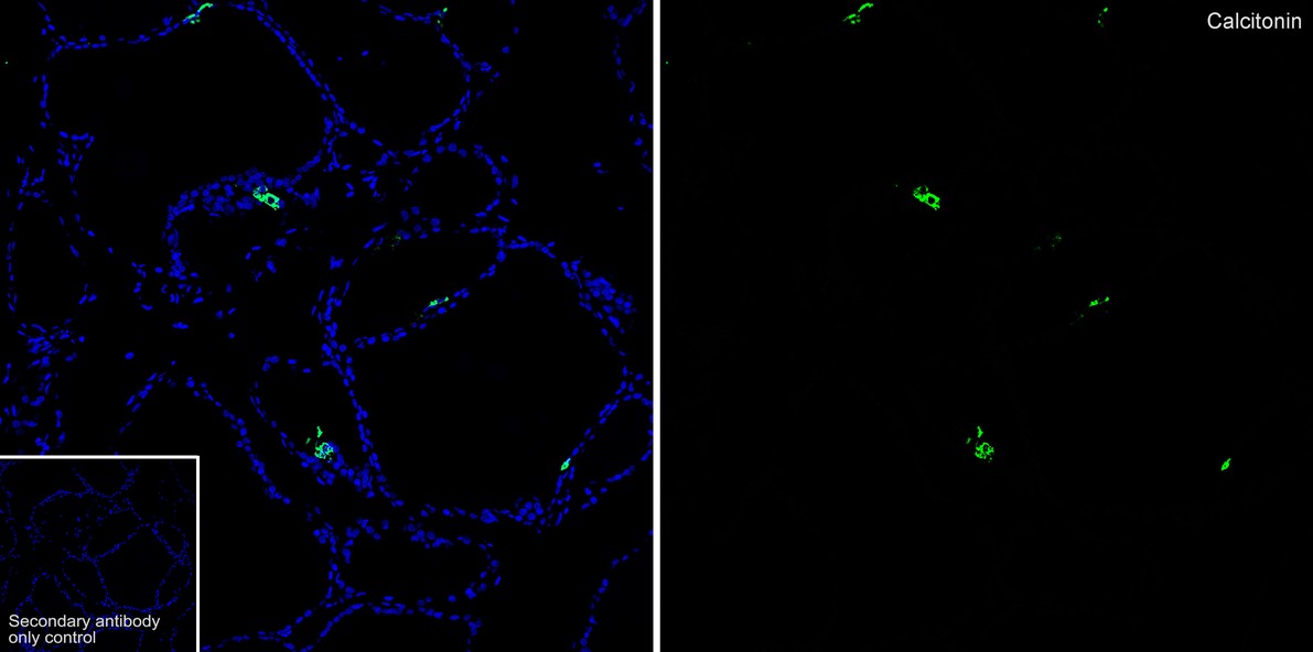
Species: Human, Site: thyroid, Sample: Paraffin-embedded section, Antibody concentration: 1/1,000.| Product Name | Calcitonin Recombinant Rabbit Monoclonal Antibody |
|---|---|
| Antibody Type | Primary Antibodies |
| Immunogen | Synthetic peptide within human Calcitonin aa 85-116. |
| Clonality | monoclonal |
|---|---|
| Isotype | IgG |
| Host Species | Rabbit |
| Tested Applications | ICC/IFIF-PIHC |
| IHC:1:2000-1:5000 ICC/IF:1:100 IF-P:1:1000 |
|
| Species Reactivity | Human |
| Concentration | 1mg/ml |
| Purification | Protein A |
| Gene Symbol | CALCA |
|---|---|
| Gene Synonyms | CT KC PCT CGRP CALC1 CGRP1 CGRP-I CGRP-alpha |
| Gene Full Name | calcitonin related polypeptide alpha |
| Gene Summary | This gene encodes the peptide hormones calcitonin, calcitonin gene-related peptide and katacalcin by tissue-specific alternative RNA splicing of the gene transcripts and cleavage of inactive precursor proteins. Calcitonin is involved in calcium regulation and acts to regulate phosphorus metabolism. Calcitonin gene-related peptide functions as a vasodilator and as an antimicrobial peptide while katacalcin is a calcium-lowering peptide. Multiple transcript variants encoding different isoforms have been found for this gene.[provided by RefSeq, Aug 2014] |
| Alternative Names | Alpha CGRP antibody alpha type CGRP antibody CALC 1 antibody CALC A antibody CALC1 antibody calcitonin 1 antibody calcitonin gene related peptide 1 antibody calcitonin gene related peptide I antibody calcitonin related polypeptide alpha antibody CGRP 1 antibody Alpha CGRP antibody alpha type CGRP antibody CALC 1 antibody CALC A antibody CALC1 antibody calcitonin 1 antibody calcitonin gene related peptide 1 antibody calcitonin gene related peptide I antibody calcitonin related polypeptide alpha antibody CGRP 1 antibody CGRP antibody CGRP I antibody CGRP1 antibody CT antibody katacalcin antibody KC antibody |
| Molecular Weight(MW) | 15 kDa |
| Cellular Localization | Secreted. |
IHC
Immunohistochemical analysis of paraffin-embedded human medullary thyroid carcinoma tissue with Rabbit anti-Calcitonin antibody at 1/20,000 dilution. The section was pre-treated using heat mediated antigen retrieval with Tris-EDTA buffer (pH 9.0) for 20 minutes. The tissues were blocked in 1% BSA for 20 minutes at room temperature, washed with ddH2O and PBS, and then probed with the primary antibody at 1/20,000 dilution for 1 hour at room temperature. The detection was performed using an HRP conjugated compact polymer system. DAB was used as the chromogen. Tissues were counterstained with hematoxylin and mounted with DPX.
IHC
Immunohistochemical analysis of paraffin-embedded human thyroid tissue with Rabbit anti-Calcitonin antibody at 1/2,000 dilution. The section was pre-treated using heat mediated antigen retrieval with Tris-EDTA buffer (pH 9.0) for 20 minutes. The tissues were blocked in 1% BSA for 20 minutes at room temperature, washed with ddH2O and PBS, and then probed with the primary antibody at 1/2,000 dilution for 1 hour at room temperature. The detection was performed using an HRP conjugated compact polymer system. DAB was used as the chromogen. Tissues were counterstained with hematoxylin and mounted with DPX.
IHC
Immunohistochemical analysis of paraffin-embedded human breast cancer tissue (negative) with Rabbit anti-Calcitonin antibody at 1/2,000 dilution. The section was pre-treated using heat mediated antigen retrieval with Tris-EDTA buffer (pH 9.0) for 20 minutes. The tissues were blocked in 1% BSA for 20 minutes at room temperature, washed with ddH2O and PBS, and then probed with the primary antibody at 1/2,000 dilution for 1 hour at room temperature. The detection was performed using an HRP conjugated compact polymer system. DAB was used as the chromogen. Tissues were counterstained with hematoxylin and mounted with DPX.
IHC
Immunohistochemical analysis of paraffin-embedded human kidney tissue (negative) with Rabbit anti-Calcitonin antibody at 1/2,000 dilution. The section was pre-treated using heat mediated antigen retrieval with Tris-EDTA buffer (pH 9.0) for 20 minutes. The tissues were blocked in 1% BSA for 20 minutes at room temperature, washed with ddH2O and PBS, and then probed with the primary antibody at 1/2,000 dilution for 1 hour at room temperature. The detection was performed using an HRP conjugated compact polymer system. DAB was used as the chromogen. Tissues were counterstained with hematoxylin and mounted with DPX.
ICC/IF
Immunocytochemistry analysis of TT cells labeling Calcitonin with Rabbit anti-Calcitonin antibody at 1/100 dilution. Cells were fixed in 4% paraformaldehyde for 20 minutes at room temperature, permeabilized with 0.1% Triton X-100 in PBS for 5 minutes at room temperature, then blocked with 1% BSA in 10% negative goat serum for 1 hour at room temperature. Cells were then incubated with Rabbit anti-Calcitonin antibody at 1/100 dilution in 1% BSA in PBST overnight at 4 ℃. Goat Anti-Rabbit IgG H&L (488) was used as the secondary antibody at 1/1,000 dilution. PBS instead of the primary antibody was used as the secondary antibody only control. Nuclear DNA was labelled in blue with DAPI. Beta tubulin (red) was stained at 1/100 dilution overnight at +4℃. Goat Anti-Mouse IgG H&L (594) was used as the secondary antibody at 1/1,000 dilution.
IF-P
Species: Human, Site: thyroid, Sample: Paraffin-embedded section, Antibody concentration: 1/1,000.| Application Notes | IHC:1:2000-1:5000 ICC/IF:1:100 IF-P:1:1000 |
|---|
| Form | Liquid |
|---|---|
| Storage Instructions | Store at +4℃ after thawing. Aliquot store at -20℃. Avoid repeated freeze / thaw cycles. |
| Storage Buffer | 1*TBS (pH7.4), 0.05% BSA, 40% Glycerol. Preservative: 0.05% Sodium Azide. |


