WB
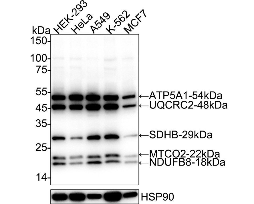
Western blot analysis of Total OXPHOS Antibody Cocktail on different lysates with Rabbit anti-Total OXPHOS Antibody Cocktail antibody at 1/2,000 dilution. Lane 1: HEK-293 cell lysate, Lane 2: HeLa cell lysate, Lane 3: A549 cell lysate, Lane 4: K-562 cell lysate, Lane 5: MCF7 cell lysate, Lysates/proteins at 20 µg/Lane. Exposure time: 15 seconds; 4-20% SDS-PAGE gel. Proteins were transferred to a PVDF membrane and blocked with 5% NFDM/TBST for 1 hour at room temperature. The primary antibody at 1/2,000 dilution was used in primary antibody dilution at 4℃ overnight. Goat Anti-Rabbit IgG - HRP Secondary Antibody at 1/50,000 dilution was used for 1 hour at room temperature.IHC
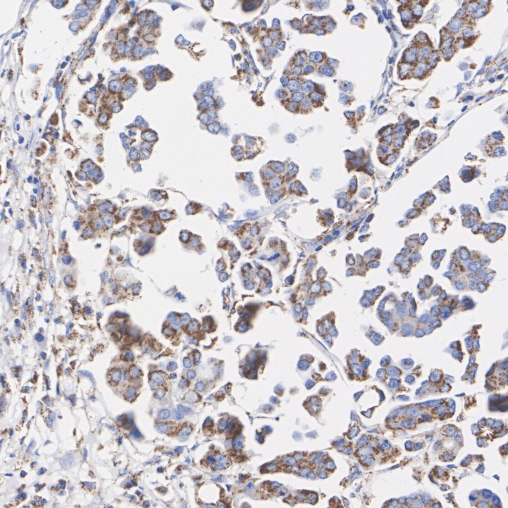
Immunohistochemical analysis of paraffin-embedded human breast carcinoma tissue with Rabbit anti-Total OXPHOS Antibody Cocktail antibody at 1/200 dilution. The section was pre-treated using heat mediated antigen retrieval with Tris-EDTA buffer (pH 9.0) for 20 minutes. The tissues were blocked in 1% BSA for 20 minutes at room temperature, washed with ddH2O and PBS, and then probed with the primary antibody at 1/200 dilution for 1 hour at room temperature. The detection was performed using an HRP conjugated compact polymer system. DAB was used as the chromogen. Tissues were counterstained with hematoxylin and mounted with DPX.IHC
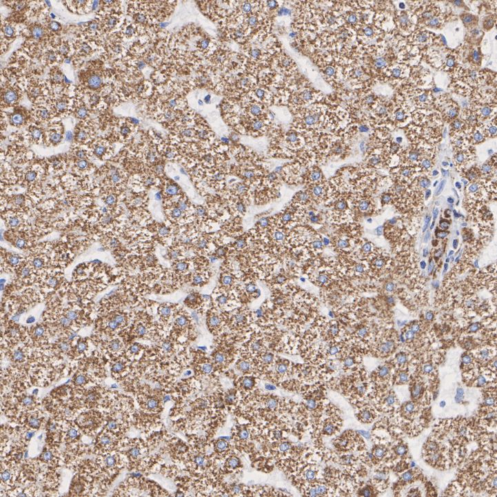
Immunohistochemical analysis of paraffin-embedded human liver tissue with Rabbit anti-Total OXPHOS Antibody Cocktail antibody at 1/200 dilution. The section was pre-treated using heat mediated antigen retrieval with Tris-EDTA buffer (pH 9.0) for 20 minutes. The tissues were blocked in 1% BSA for 20 minutes at room temperature, washed with ddH2O and PBS, and then probed with the primary antibody at 1/200 dilution for 1 hour at room temperature. The detection was performed using an HRP conjugated compact polymer system. DAB was used as the chromogen. Tissues were counterstained with hematoxylin and mounted with DPX.| Product Name | Total OXPHOS Antibody Cocktail Recombinant Rabbit Multiclonal Antibody |
|---|---|
| Antibody Type | Primary Antibodies |
| Clonality | Monoclonal |
|---|---|
| Isotype | IgG |
| Host Species | Rabbit |
| Tested Applications | IHCWB |
| WB:1:2000 IHC:1:200 |
|
| Species Reactivity | Human |
| Concentration | 1mg/ml |
| Purification | Protein A |
| Alternative Names | ASHI antibody ATP synthase subunit alpha antibody Atp5a1 antibody ATP5AL2 antibody COII antibody Complex I ASHI subunit antibody COX 2 antibody COX II antibody COX2 antibody COXII antibody ASHI antibody ATP synthase subunit alpha antibody Atp5a1 antibody ATP5AL2 antibody COII antibody Complex I ASHI subunit antibody COX 2 antibody COX II antibody COX2 antibody COXII antibody CWS2 antibody Cytochrome b c1 complex subunit 2 mitochondrial antibody Cytochrome c oxidase II antibody EC 1.10.2.2 antibody FLJ92337 antibody Iron sulfur subunit antibody Iron sulfur subunit of complex II antibody MC3DN5 antibody MC5DN4 antibody mitochondrial antibody MT CO2 antibody MTCO2 antibody NADH dehydrogenase (ubiquinone) 1 beta subcomplex 8 19kDa antibody NADH dehydrogenase [ubiquinone] 1 beta subcomplex subunit 8 mitochondrial antibody PGL 4 antibody PGL4 antibody QCR2 antibody SDH 1 antibody SDH1 antibody SDH2 antibody SDH2 homolog of antibody SDHIP antibody Succinate dehydrogenase [ubiquinone] iron sulfur protein mitochondrial antibody Succinate dehydrogenase [ubiquinone] iron sulfur subunit antibody succinate dehydrogenase [ubiquinone] iron-sulfur subunit mitochondrial antibody Succinate Dehydrogenase 1 Iron Sulfur Subunit antibody Succinate Dehydrogenase 2 S. cerevisiae homolog of antibody Succinate dehydrogenase complex iron sulfur subunit B antibody Succinate dehydrogenase complex subunit B iron sulfur antibody Ubiquinol cytochrome c reductase complex core protein 2 mitochondrial antibody Ubiquinol cytochrome c reductase core protein II antibody UQCR2 antibody |
|---|---|
| Molecular Weight(MW) | Predicted band size: 18/22/29/48/54 kDa |
| Cellular Localization | Mitochondrion inner membrane. |
WB
Western blot analysis of Total OXPHOS Antibody Cocktail on different lysates with Rabbit anti-Total OXPHOS Antibody Cocktail antibody at 1/2,000 dilution. Lane 1: HEK-293 cell lysate, Lane 2: HeLa cell lysate, Lane 3: A549 cell lysate, Lane 4: K-562 cell lysate, Lane 5: MCF7 cell lysate, Lysates/proteins at 20 µg/Lane. Exposure time: 15 seconds; 4-20% SDS-PAGE gel. Proteins were transferred to a PVDF membrane and blocked with 5% NFDM/TBST for 1 hour at room temperature. The primary antibody at 1/2,000 dilution was used in primary antibody dilution at 4℃ overnight. Goat Anti-Rabbit IgG - HRP Secondary Antibody at 1/50,000 dilution was used for 1 hour at room temperature.
IHC
Immunohistochemical analysis of paraffin-embedded human breast carcinoma tissue with Rabbit anti-Total OXPHOS Antibody Cocktail antibody at 1/200 dilution. The section was pre-treated using heat mediated antigen retrieval with Tris-EDTA buffer (pH 9.0) for 20 minutes. The tissues were blocked in 1% BSA for 20 minutes at room temperature, washed with ddH2O and PBS, and then probed with the primary antibody at 1/200 dilution for 1 hour at room temperature. The detection was performed using an HRP conjugated compact polymer system. DAB was used as the chromogen. Tissues were counterstained with hematoxylin and mounted with DPX.
IHC
Immunohistochemical analysis of paraffin-embedded human liver tissue with Rabbit anti-Total OXPHOS Antibody Cocktail antibody at 1/200 dilution. The section was pre-treated using heat mediated antigen retrieval with Tris-EDTA buffer (pH 9.0) for 20 minutes. The tissues were blocked in 1% BSA for 20 minutes at room temperature, washed with ddH2O and PBS, and then probed with the primary antibody at 1/200 dilution for 1 hour at room temperature. The detection was performed using an HRP conjugated compact polymer system. DAB was used as the chromogen. Tissues were counterstained with hematoxylin and mounted with DPX.| Application Notes | WB:1:2000 IHC:1:200 |
|---|
| Form | Liquid |
|---|---|
| Storage Instructions | Store at +4℃ after thawing. Aliquot store at -20℃. Avoid repeated freeze / thaw cycles. |
| Storage Buffer | PBS (pH7.4), 0.1% BSA, 40% Glycerol. Preservative: 0.05% Sodium Azide. |


