WB
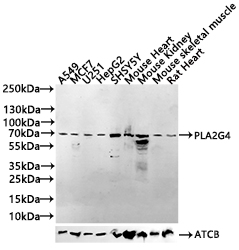
Positive WB detected in: A549 whole cell lysate(30µg), MCF7 whole cell lysate(30µg), U251 whole cell lysate(320µg), HepG2 whole cell lysate(30µg), SHSY5Y whole cell lysate(30µg), Mouse Heart tissue lysate30µg),Mouse Kidney tissue lysate30µg),Mouse skeletal muscle tissue lysate30µg),Rat Heart tissue lysate30µg), All lanes: PLA2G4C antibody at 1:500 Secondary Goat polyclonal to rabbit IgG at 1/40000 dilution. Exposure time: 120s.IHC
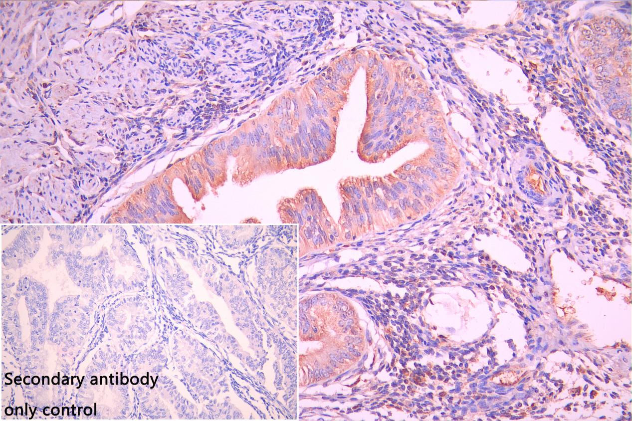
IHC image of PLA2G4C Antibody diluted at 1:50 and staining in paraffin-embedded human endometrial cancer performed on a Leica BondTM system. After dewaxing and hydration, antigen retrieval was mediated by high pressure in a citrate buffer (pH 6.0). Section was blocked with 10% normal goat serum 30min at RT. Then primary antibody (1% BSA) was incubated at 4°C overnight. The primary is detected by a Goat anti-rabbit polymer IgG labeled by HRP and visualized using 0.05% DAB. Secondary antibody only control: uses 1% BSA instead of primary antibody.ICC/IF
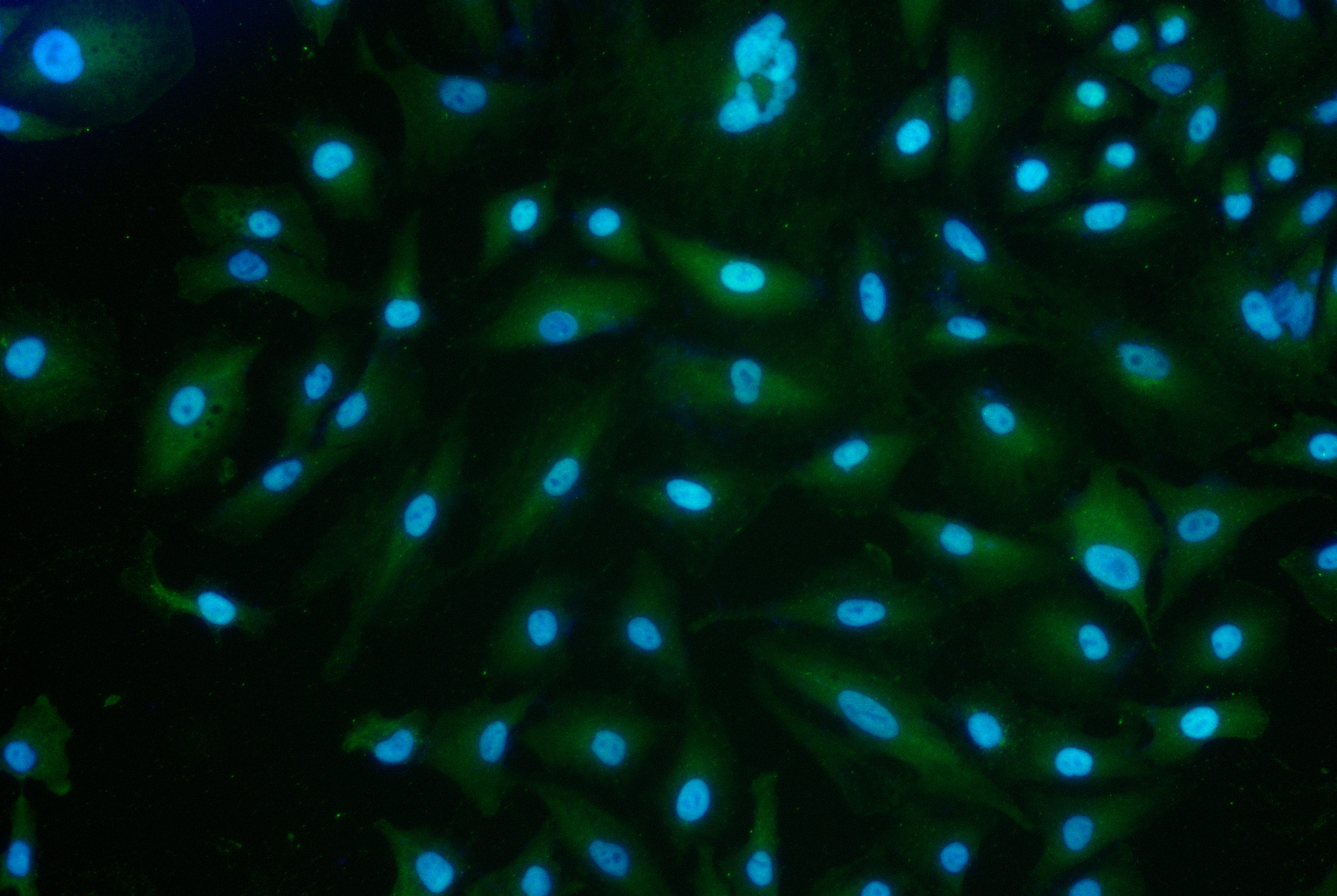
Immunofluorescence staining of A549 cell with PLA2G4C Antibody at 1:30 counter-stained with DAPI. The cells were fixed in 4% formaldehyde and and permeated by 0.2% TritonX-100 for 15 min. Then 10% normal goat serum to block non-specific protein-protein interactions . The cells were then incubated with the antibody overnight at 4℃. The secondary antibody was Alexa Fluor 488-congugated AffiniPure Goat Anti-Rabbit IgG(H+L).| Product Name | Rabbit anti-PLA2G4C Polyclonal antibody |
|---|---|
| Antibody Type | Primary Antibodies |
| Immunogen | synthesized peptide derived from human PLA2G4C |
| Clonality | Polyclonal |
|---|---|
| Isotype | IgG |
| Host Species | Rabbit |
| Tested Applications | ICC/IFIHCWB |
| WB:1:500-1:2000 IHC:1:20-1:100 ICC/IF:1:20-1:100 |
|
| Species Reactivity | HumanMouseRat |
| Concentration | 1mg/ml |
| Purification | Affinity purified |
| Gene Symbol | PLA2G4C |
|---|---|
| Gene Synonyms | CPLA2-gamma |
| Gene Full Name | phospholipase A2 group IVC |
| Gene Summary | This gene encodes a protein which is a member of the phospholipase A2 enzyme family which hydrolyzes glycerophospholipids to produce free fatty acids and lysophospholipids, both of which serve as precursors in the production of signaling molecules. The encoded protein has been shown to be a calcium-independent and membrane bound enzyme. Multiple transcript variants encoding different isoforms have been found for this gene. [provided by RefSeq, Apr 2009] |
| Alternative Names | cPLA2 gamma antibody Cytosolic phospholipase A2 gamma antibody EC 3.1.1.4 antibody PA24C_HUMAN antibody Phospholipase A2 group IVC antibody PLA2G4C antibody |
| Molecular Weight(MW) | 70 kDa |
| Cellular Localization | Cell membrane; Lipid-anchor. Endoplasmic reticulum membrane; Lipid-anchor. Mitochondrion membrane; Lipid-anchor. Lipid droplet. |
WB
Positive WB detected in: A549 whole cell lysate(30µg), MCF7 whole cell lysate(30µg), U251 whole cell lysate(320µg), HepG2 whole cell lysate(30µg), SHSY5Y whole cell lysate(30µg), Mouse Heart tissue lysate30µg),Mouse Kidney tissue lysate30µg),Mouse skeletal muscle tissue lysate30µg),Rat Heart tissue lysate30µg), All lanes: PLA2G4C antibody at 1:500 Secondary Goat polyclonal to rabbit IgG at 1/40000 dilution. Exposure time: 120s.
IHC
IHC image of PLA2G4C Antibody diluted at 1:50 and staining in paraffin-embedded human endometrial cancer performed on a Leica BondTM system. After dewaxing and hydration, antigen retrieval was mediated by high pressure in a citrate buffer (pH 6.0). Section was blocked with 10% normal goat serum 30min at RT. Then primary antibody (1% BSA) was incubated at 4°C overnight. The primary is detected by a Goat anti-rabbit polymer IgG labeled by HRP and visualized using 0.05% DAB. Secondary antibody only control: uses 1% BSA instead of primary antibody.
ICC/IF
Immunofluorescence staining of A549 cell with PLA2G4C Antibody at 1:30 counter-stained with DAPI. The cells were fixed in 4% formaldehyde and and permeated by 0.2% TritonX-100 for 15 min. Then 10% normal goat serum to block non-specific protein-protein interactions . The cells were then incubated with the antibody overnight at 4℃. The secondary antibody was Alexa Fluor 488-congugated AffiniPure Goat Anti-Rabbit IgG(H+L).| Application Notes | WB:1:500-1:2000 IHC:1:20-1:100 ICC/IF:1:20-1:100 |
|---|
| Form | Liquid |
|---|---|
| Storage Instructions | Upon receipt, store at -20°C or -80°C. Avoid repeated freeze. |
| Storage Buffer | 0.03% Proclin 300 Constituents: 50% Glycerol, 0.01M PBS, PH 7.4 |


