WB
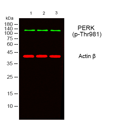
Western blot analysis of lysates from 1) 453, 2) AD293 , 3) Hela cells, (Green) primary antibody was diluted at 1:1000, 4°over night, secondary antibody was diluted at 1:10000, 37° 1hour. (Red) Actin β Monoclonal Antibody antibody was diluted at 1:5000 as loading control, 4° over night,secondary antibody was diluted at 1:10000, 37° 1hour.IHC
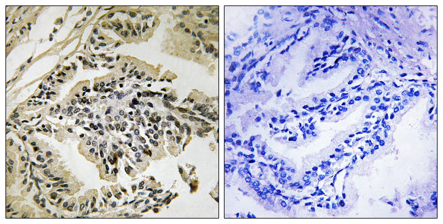
Immunohistochemistry analysis of paraffin-embedded human prostate carcinoma, using PEK/PERK (Phospho-Thr981) Antibody. The picture on the right is blocked with the phospho peptide.IHC
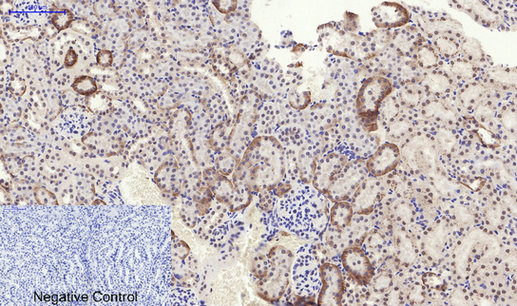
Immunohistochemical analysis of paraffin-embedded Mouse-kidney tissue. 1,PERK (phospho Thr981) Polyclonal Antibody was diluted at 1:200(4℃,overnight). 2, Sodium citrate pH 6.0 was used for antibody retrieval(>98℃,20min). 3,Secondary antibody was diluted at 1:200(room tempeRature, 30min). Negative control was used by secondary antibody only.IHC
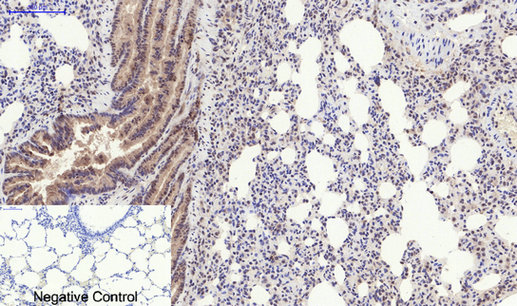
Immunohistochemical analysis of paraffin-embedded Rat-lung tissue. 1,PERK (phospho Thr981) Polyclonal Antibody was diluted at 1:200(4℃,overnight). 2, Sodium citrate pH 6.0 was used for antibody retrieval(>98℃,20min). 3,Secondary antibody was diluted at 1:200(room tempeRature, 30min). Negative control was used by secondary antibody only.ICC/IF
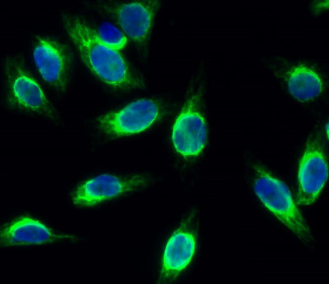
Immunofluorescence analysis of Hela cell. 1,PERK (phospho Thr981) Polyclonal Antibody(green) was diluted at 1:200(4° overnight). 2, Goat Anti Rabbit Alexa Fluor 488 was diluted at 1:1000(room temperature, 50min). 3 DAPI(blue) 10min.| Product Name | PERK (Phospho Thr982) Rabbit pAb |
|---|---|
| Antibody Type | Primary Antibodies |
| Immunogen | The antiserum was produced against synthesized peptide derived from human PEK/PERK around the phosphorylation site of Thr982. AA range:947-996. |
| Modification | p-Thr982 |
| Clonality | Polyclonal |
|---|---|
| Isotype | IgG |
| Host Species | Rabbit |
| Tested Applications | ICC/IFIHCWB |
| WB:1:500-1:2000 IHC:1:100-1:300 ICC/IF:1:50-1:200 |
|
| Species Reactivity | Human |
| Concentration | 1mg/ml |
| Purification | Protein A |
| Gene Symbol | EIF2AK3 |
|---|---|
| Gene Synonyms | PEK WRS PERK |
| Gene Full Name | eukaryotic translation initiation factor 2 alpha kinase 3 |
| Gene Summary | The protein encoded by this gene phosphorylates the alpha subunit of eukaryotic translation-initiation factor 2, leading to its inactivation, and thus to a rapid reduction of translational initiation and repression of global protein synthesis. This protein is thought to modulate mitochondrial function. It is a type I membrane protein located in the endoplasmic reticulum (ER), where it is induced by ER stress caused by malfolded proteins. Mutations in this gene are associated with Wolcott-Rallison syndrome. [provided by RefSeq, Sep 2015] |
| Alternative Names | EIF2AK3 PEK PERK Eukaryotic translation initiation factor 2-alpha kinase 3 PRKR-like endoplasmic reticulum kinase Pancreatic eIF2-alpha kinase HsPEK |
| Molecular Weight(MW) | 130kDa |
| Cellular Localization | Endoplasmic reticulum membrane; Single-pass type I membrane protein. |
WB
Western blot analysis of lysates from 1) 453, 2) AD293 , 3) Hela cells, (Green) primary antibody was diluted at 1:1000, 4°over night, secondary antibody was diluted at 1:10000, 37° 1hour. (Red) Actin β Monoclonal Antibody antibody was diluted at 1:5000 as loading control, 4° over night,secondary antibody was diluted at 1:10000, 37° 1hour.
IHC
Immunohistochemistry analysis of paraffin-embedded human prostate carcinoma, using PEK/PERK (Phospho-Thr981) Antibody. The picture on the right is blocked with the phospho peptide.
IHC
Immunohistochemical analysis of paraffin-embedded Mouse-kidney tissue. 1,PERK (phospho Thr981) Polyclonal Antibody was diluted at 1:200(4℃,overnight). 2, Sodium citrate pH 6.0 was used for antibody retrieval(>98℃,20min). 3,Secondary antibody was diluted at 1:200(room tempeRature, 30min). Negative control was used by secondary antibody only.
IHC
Immunohistochemical analysis of paraffin-embedded Rat-lung tissue. 1,PERK (phospho Thr981) Polyclonal Antibody was diluted at 1:200(4℃,overnight). 2, Sodium citrate pH 6.0 was used for antibody retrieval(>98℃,20min). 3,Secondary antibody was diluted at 1:200(room tempeRature, 30min). Negative control was used by secondary antibody only.
ICC/IF
Immunofluorescence analysis of Hela cell. 1,PERK (phospho Thr981) Polyclonal Antibody(green) was diluted at 1:200(4° overnight). 2, Goat Anti Rabbit Alexa Fluor 488 was diluted at 1:1000(room temperature, 50min). 3 DAPI(blue) 10min.| Application Notes | WB:1:500-1:2000 IHC:1:100-1:300 ICC/IF:1:50-1:200 |
|---|
| Form | Liquid |
|---|---|
| Storage Instructions | -15°C to -25°C/1 year(Do not lower than -25°C) |
| Storage Buffer | PBS, 50% glycerol, 0.05% Proclin 300, 0.05%BSA. |


