WB
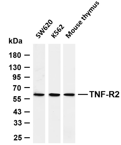
Various whole cell lysates were separated by 4-20% SDS-PAGE, and the membrane was blotted with anti-TNF-R2 antibody. The HRP-conjugated Goat anti-Rabbit IgG (H + L) antibody was used to detect the antibody. Lane 1: SW620 Lane 2: K562 Lane 3: Mouse thymus Predicted band size: 48kDa Observed band size: 60kDaIF/ICC
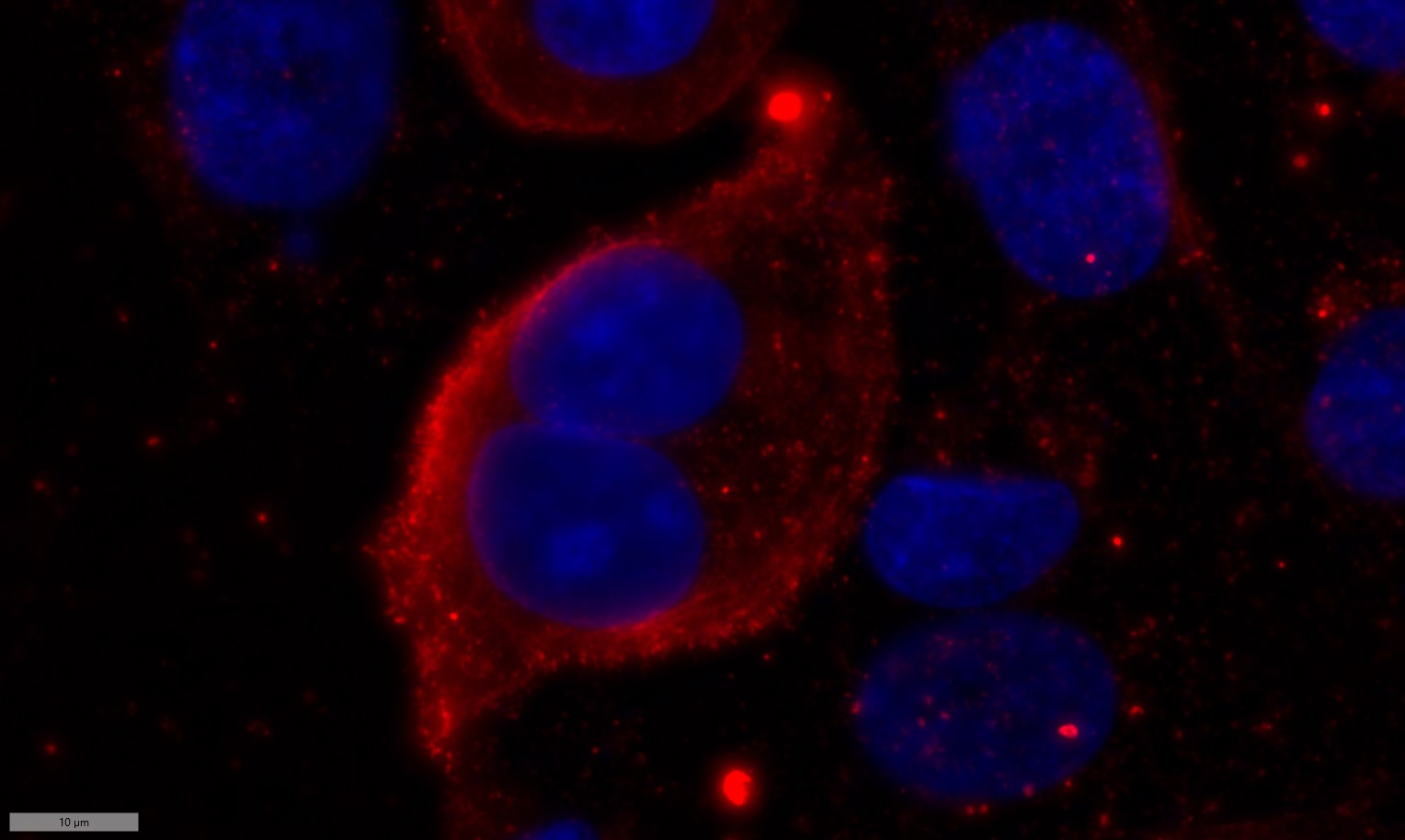
Immunofluorescence analysis of Siha cell. 1,primary Antibody was diluted at 1:100(4℃ overnight). 2, Goat Anti Rabbit IgG (H&L) - AFluor 594 Secondary antibody was diluted at 1:500(room temperature, 50min).| Product Name | TNF-R2 Rabbit mAb |
|---|---|
| Antibody Type | Primary Antibodies |
| Immunogen | Tumor necrosis factor receptor superfamily member 1B |
| Clonality | Polyclonal |
|---|---|
| Isotype | IgG |
| Host Species | Rabbit |
| Tested Applications | ELISAIFIHCIPWB |
| IHC :200-1:1000 WB:2000-1:10000 IF:200-1:1000 ELISA:5000-1:20000 IP:50-1:200 |
|
| Species Reactivity | HumanMouseRat |
| Concentration | 1μg/μl |
| Purification | Protein A |
| Gene Symbol | TNFRSF1B |
|---|---|
| Gene Synonyms | TNFRSF1B TNFBR TNFR2 |
| Gene Full Name | Tumor necrosis factor receptor superfamily member 1B |
| Gene Summary | The protein encoded by this gene is a member of the TNF-receptor superfamily. This protein and TNF-receptor 1 form a heterocomplex that mediates the recruitment of two anti-apoptotic proteins, c-IAP1 and c-IAP2, which possess E3 ubiquitin ligase activity. The function of IAPs in TNF-receptor signalling is unknown, however, c-IAP1 is thought to potentiate TNF-induced apoptosis by the ubiquitination and degradation of TNF-receptor-associated factor 2, which mediates anti-apoptotic signals. Knockout studies in mice also suggest a role of this protein in protecting neurons from apoptosis by stimulating antioxidative pathways. |
| Alternative Names | TNFRSF1B TNFBR TNFR2 Tumor necrosis factor receptor superfamily member 1B Tumor necrosis factor receptor 2 TNF-R2 Tumor necrosis factor receptor type II TNF-RII TNFR-II p75 p80 TNF-alpha receptor CD antigen CD120b Etanercept |
| Molecular Weight(MW) | 48kd |
| SwissProt ID | P20333,P25119,Q80WY6 |
|---|
WB
Various whole cell lysates were separated by 4-20% SDS-PAGE, and the membrane was blotted with anti-TNF-R2 antibody. The HRP-conjugated Goat anti-Rabbit IgG (H + L) antibody was used to detect the antibody. Lane 1: SW620 Lane 2: K562 Lane 3: Mouse thymus Predicted band size: 48kDa Observed band size: 60kDa
IF/ICC
Immunofluorescence analysis of Siha cell. 1,primary Antibody was diluted at 1:100(4℃ overnight). 2, Goat Anti Rabbit IgG (H&L) - AFluor 594 Secondary antibody was diluted at 1:500(room temperature, 50min).| Application Notes | IHC :200-1:1000 WB:2000-1:10000 IF:200-1:1000 ELISA:5000-1:20000 IP:50-1:200 |
|---|
| Form | Liquid |
|---|---|
| Storage Instructions | -15°C to -25°C/1 year(Do not lower than -25°C) |
| Storage Buffer | PBS, 50% glycerol, 0.05% Proclin 300, 0.05%BSA |


