WB
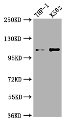
Positive WB detected in: THP-1 whole cell lysate, K562 whole cell lysate, All lanes: C3 antibody at 1:1000, Secondary Goat polyclonal to rabbit IgG at 1/50000 dilution.IHC
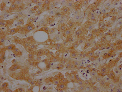
IHC image ofC3 Recombinant Monoclonal Antibody diluted at 1:100 and staining in paraffin-embedded human liver tissue performed on a Leica BondTM system. After dewaxing and hydration, antigen retrieval was mediated by high pressure in a citrate buffer (pH 6.0). Section was blocked with 10% normal goat serum 30min at RT. Then primary antibody (1% BSA) was incubated at 4℃ overnight. The primary is detected by a Goat anti-rabbit IgG polymer labeled by HRP and visualized using 0.05% DAB.FC
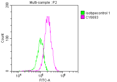
Overlay histogram showing HepG2 cells stained withC3 Recombinant Monoclonal Antibody (red line) at 1:50. The cells were fixed with 70% Ethylalcohol (18h) and then incubated in 10% normal goat serum to block non-specific protein-protein interactions followedby the antibody (1µg/1*106 cells) for 1 h at 4℃.The secondary antibody used was FITC-conjugated goat anti-rabbit IgG (H+L) at 1/200 dilution for 30min at 4℃. Control antibody (green line) was Rabbit IgG (1µg/1*106 cells) used under the same conditions. Acquisition of >10,000 events was performed.| Product Name | C3 Recombinant Monoclonal Antibody |
|---|---|
| Antibody Type | Primary Antibodies |
| Immunogen | A synthesized peptide derived from human C3 |
| Clonality | Monoclonal |
|---|---|
| Isotype | IgG |
| Host Species | Rabbit |
| Tested Applications | FCIHCWB |
| WB:1:500-1:2000 IHC:1:50-1:200 FC:1:20-1:200 |
|
| Species Reactivity | Human |
| Concentration | 1mg/ml |
| Purification | Affinity purified |
| Gene Symbol | C3 |
|---|---|
| Gene Synonyms | ASP C3a C3b AHUS5 ARMD9 CPAMD1 HEL-S-62p |
| Gene Full Name | complement C3 |
| Gene Summary | Complement component C3 plays a central role in the activation of complement system. Its activation is required for both classical and alternative complement activation pathways. The encoded preproprotein is proteolytically processed to generate alpha and beta subunits that form the mature protein, which is then further processed to generate numerous peptide products. The C3a peptide, also known as the C3a anaphylatoxin, modulates inflammation and possesses antimicrobial activity. Mutations in this gene are associated with atypical hemolytic uremic syndrome and age-related macular degeneration in human patients. [provided by RefSeq, Nov 2015] |
| Alternative Names | Complement C3 (C3 and PZP-like alpha-2-macroglobulin domain-containing protein 1) [Cleaved into: Complement C3 beta chain C3-beta-c (C3bc) Complement C3 alpha chain C3a anaphylatoxin Acylation stimulating protein (ASP) (C3adesArg) Complement C3b alpha' chain Complement C3c alpha' chain fragment 1 Complement C3dg fragment Complement C3g fragment Complement C3d fragment Complement C3f fragment Complement C3c alpha' chain fragment 2] C3 CPAMD1 |
| Molecular Weight(MW) | 188 kDa(Observed band size: 115 kDa) |
| Cellular Localization | Secreted. |
WB
Positive WB detected in: THP-1 whole cell lysate, K562 whole cell lysate, All lanes: C3 antibody at 1:1000, Secondary Goat polyclonal to rabbit IgG at 1/50000 dilution.
IHC
IHC image ofC3 Recombinant Monoclonal Antibody diluted at 1:100 and staining in paraffin-embedded human liver tissue performed on a Leica BondTM system. After dewaxing and hydration, antigen retrieval was mediated by high pressure in a citrate buffer (pH 6.0). Section was blocked with 10% normal goat serum 30min at RT. Then primary antibody (1% BSA) was incubated at 4℃ overnight. The primary is detected by a Goat anti-rabbit IgG polymer labeled by HRP and visualized using 0.05% DAB.
FC
Overlay histogram showing HepG2 cells stained withC3 Recombinant Monoclonal Antibody (red line) at 1:50. The cells were fixed with 70% Ethylalcohol (18h) and then incubated in 10% normal goat serum to block non-specific protein-protein interactions followedby the antibody (1µg/1*106 cells) for 1 h at 4℃.The secondary antibody used was FITC-conjugated goat anti-rabbit IgG (H+L) at 1/200 dilution for 30min at 4℃. Control antibody (green line) was Rabbit IgG (1µg/1*106 cells) used under the same conditions. Acquisition of >10,000 events was performed.| Application Notes | WB:1:500-1:2000 IHC:1:50-1:200 FC:1:20-1:200 |
|---|
| Form | Liquid |
|---|---|
| Storage Instructions | Upon receipt, store at -20°C or -80°C. Avoid repeated freeze. |
| Storage Buffer | 0.03% Proclin 300 Constituents: 50% Glycerol, 0.01M PBS, PH 7.4 |


