IHC
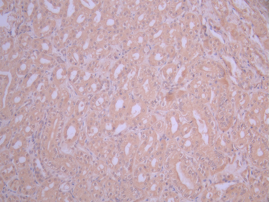
IHC image of ACKR1 Recombinant Monoclonal Antibody diluted at 1:50 and staining in paraffin-embedded human kidney tissue performed on a Leica BondTM system. After dewaxing and hydration, antigen retrieval was mediated by high pressure in a citrate buffer (pH 6.0). Section was blocked with 10% normal goat serum 30min at RT. Then primary antibody (1% BSA) was incubated at 4°C overnight. The primary is detected by a Anti-Human lgG, Fcy Fragment Specific labeled by HRP and visualized using 0.05% DAB.ICC/IF
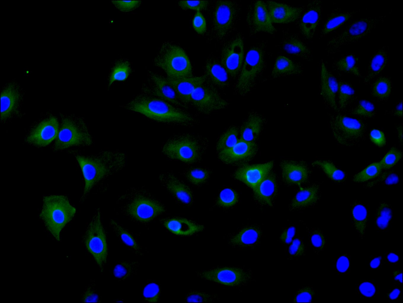
Immunofluorescence staining of A549 cell with ACKR1 Recombinant Monoclonal Antibody at 1:30 ,counter-stained with DAPI. The cells were fixed in 4% formaldehyde, permeabilized using 0.2% Triton X-100 and blocked in 10% normal Goat Serum. The cells were then incubated with the antibody overnight at 4°C. The secondary antibody was Fluorescein (FITC) AffiniPure Goat Anti-Human IgG, Fcγ fragment specific.| Product Name | ACKR1 Recombinant Monoclonal Antibody |
|---|---|
| Antibody Type | Primary Antibodies |
| Immunogen | Recombinant Human ACKR1 protein |
| Clonality | Monoclonal |
|---|---|
| Isotype | IgG1 |
| Tested Applications | ICC/IFIHC |
| IHC:1:50-1:200 ICC/IF:1:50-1:200 |
|
| Species Reactivity | Human |
| Concentration | 1mg/ml |
| Purification | Affinity purified |
| Gene Symbol | ACKR1 |
|---|---|
| Gene Synonyms | FY Dfy GPD DARC GpFy CCBP1 CD234 WBCQ1 DARC/ACKR1 |
| Gene Full Name | atypical chemokine receptor 1 (Duffy blood group) |
| Gene Summary | The protein encoded by this gene is a glycosylated membrane protein and a non-specific receptor for several chemokines. The encoded protein is the receptor for the human malarial parasites Plasmodium vivax and Plasmodium knowlesi. Polymorphisms in this gene are the basis of the Duffy blood group system. Two transcript variants encoding different isoforms have been found for this gene. [provided by RefSeq, Jul 2008] |
| Alternative Names | ACKR1 antibody Atypical chemokine receptor 1 antibody CCBP1 antibody CD234 antibody DARC antibody Dfy antibody Duffy antigen/chemokine receptor antibody duffy blood group antibody Duffy blood group antigen antibody Duffy blood group chemokine receptor antibody DUFFY_HUMAN antibody FY antibody Fy glycoprotein antibody Glycoprotein D antibody GPD antibody GpFy antibody Plasmodium vivax receptor antibody WBCQ1 antibody |
| Cellular Localization | Early endosome. Recycling endosome. Membrane; Multi-pass membrane protein. Note=Predominantly localizes to endocytic vesicles, and upon stimulation by the ligand is internalized via caveolae. Once internalized, the ligand dissociates from the receptor, and is targeted to degradation while the receptor is recycled back to the cell membrane. |
IHC
IHC image of ACKR1 Recombinant Monoclonal Antibody diluted at 1:50 and staining in paraffin-embedded human kidney tissue performed on a Leica BondTM system. After dewaxing and hydration, antigen retrieval was mediated by high pressure in a citrate buffer (pH 6.0). Section was blocked with 10% normal goat serum 30min at RT. Then primary antibody (1% BSA) was incubated at 4°C overnight. The primary is detected by a Anti-Human lgG, Fcy Fragment Specific labeled by HRP and visualized using 0.05% DAB.
ICC/IF
Immunofluorescence staining of A549 cell with ACKR1 Recombinant Monoclonal Antibody at 1:30 ,counter-stained with DAPI. The cells were fixed in 4% formaldehyde, permeabilized using 0.2% Triton X-100 and blocked in 10% normal Goat Serum. The cells were then incubated with the antibody overnight at 4°C. The secondary antibody was Fluorescein (FITC) AffiniPure Goat Anti-Human IgG, Fcγ fragment specific.| Application Notes | IHC:1:50-1:200 ICC/IF:1:50-1:200 |
|---|
| Form | Liquid |
|---|---|
| Storage Instructions | Upon receipt, store at -20°C or -80°C. Avoid repeated freeze. |
| Storage Buffer | 0.03% Proclin 300 Constituents: 50% Glycerol, 0.01M PBS, PH 7.4 |


