WB
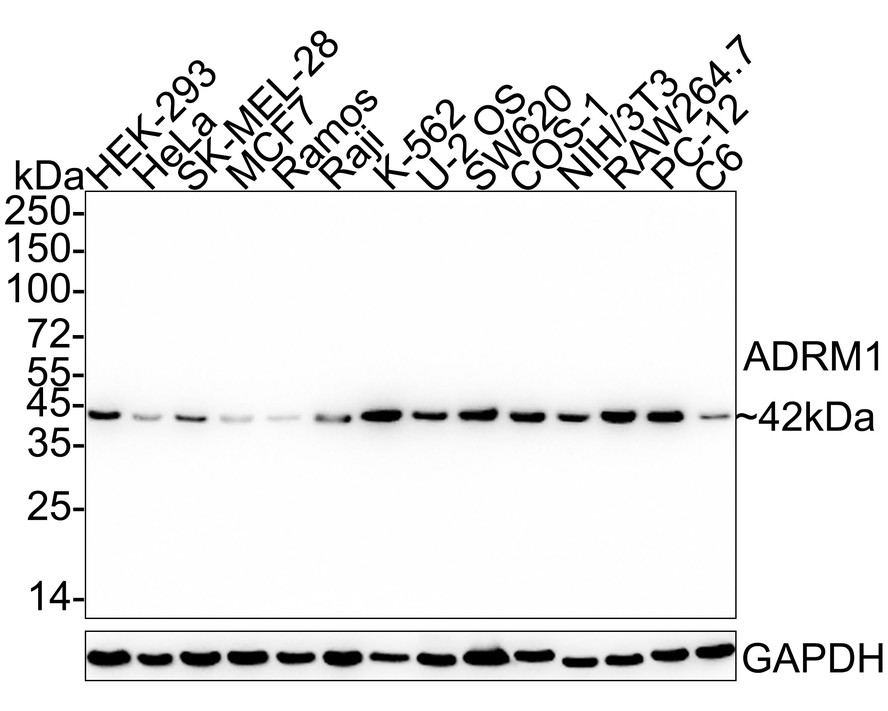
Western blot analysis of ADRM1 on different lysates with Rabbit anti-ADRM1 antibody at 1/2,000 dilution. Lane 1: HEK-293 cell lysate (20 µg/Lane), Lane 2: HeLa cell lysate (20 µg/Lane), Lane 3: SK-MEL-28 cell lysate (20 µg/Lane), Lane 4: MCF7 cell lysate (20 µg/Lane), Lane 5: Ramos cell lysate (20 µg/Lane), Lane 6: Raji cell lysate (20 µg/Lane), Lane 7: K-562 cell lysate (20 µg/Lane), Lane 8: U-2 OS cell lysate (20 µg/Lane), Lane 9: SW620 cell lysate (20 µg/Lane), Lane 10: COS-1 cell lysate (20 µg/Lane), Lane 11: NIH/3T3 cell lysate (20 µg/Lane), Lane 12: RAW264.7 cell lysate (20 µg/Lane), Lane 13: PC-12 cell lysate (20 µg/Lane), Lane 14: C6 cell lysate (20 µg/Lane), Exposure time: 3 minutes; 4-20% SDS-PAGE gel. Proteins were transferred to a PVDF membrane and blocked with 5% NFDM/TBST for 1 hour at room temperature. The primary antibody at 1/2,000 dilution was used in 5% NFDM/TBST at 4℃ overnight. Goat Anti-Rabbit IgG - HRP Secondary Antibody at 1/50,000 dilution was used for 1 hour at room temperature.ICC/IF
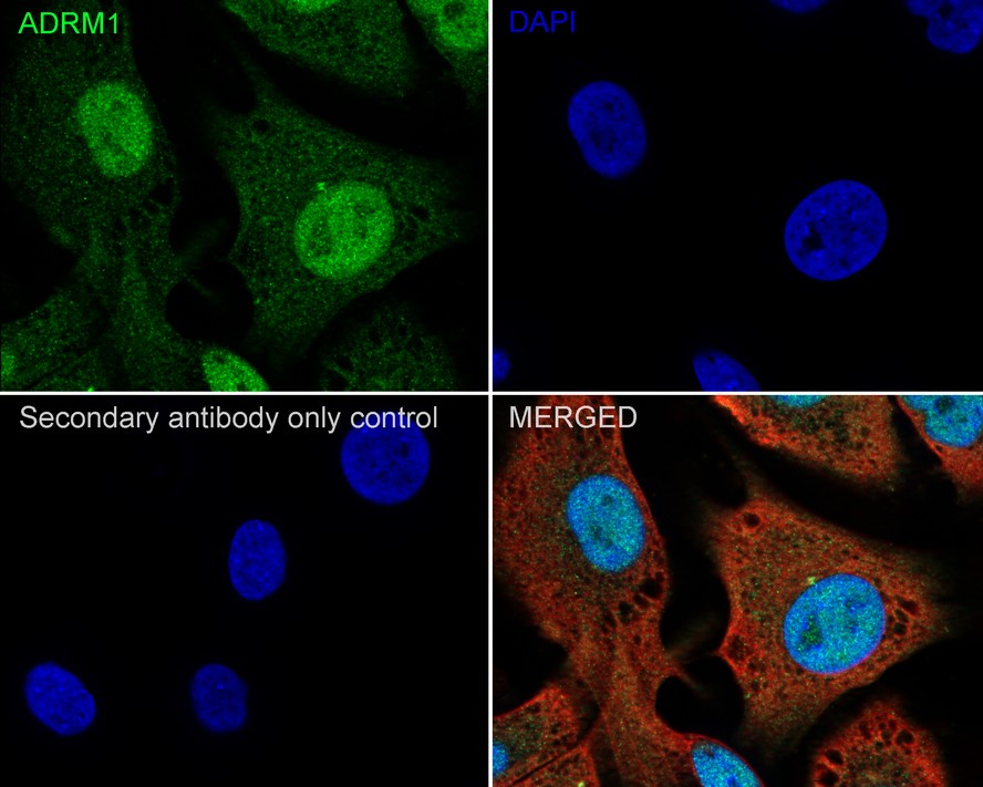
Immunocytochemistry analysis of SK-MEL-28 cells labeling ADRM1 with Rabbit anti-ADRM1 antibody at 1/500 dilution. Cells were fixed in 4% paraformaldehyde for 20 minutes at room temperature, permeabilized with 0.1% Triton X-100 in PBS for 5 minutes at room temperature, then blocked with 1% BSA in 10% negative goat serum for 1 hour at room temperature. Cells were then incubated with Rabbit anti-ADRM1 antibody at 1/500 dilution in 1% BSA in PBST overnight at 4 ℃. Goat Anti-Rabbit IgG H&L (488) was used as the secondary antibody at 1/1,000 dilution. PBS instead of the primary antibody was used as the secondary antibody only control. Nuclear DNA was labelled in blue with DAPI. Beta tubulin (red) was stained at 1/100 dilution overnight at +4℃. Goat Anti-Mouse IgG H&L (594) was used as the secondary antibody at 1/1,000 dilution.FC
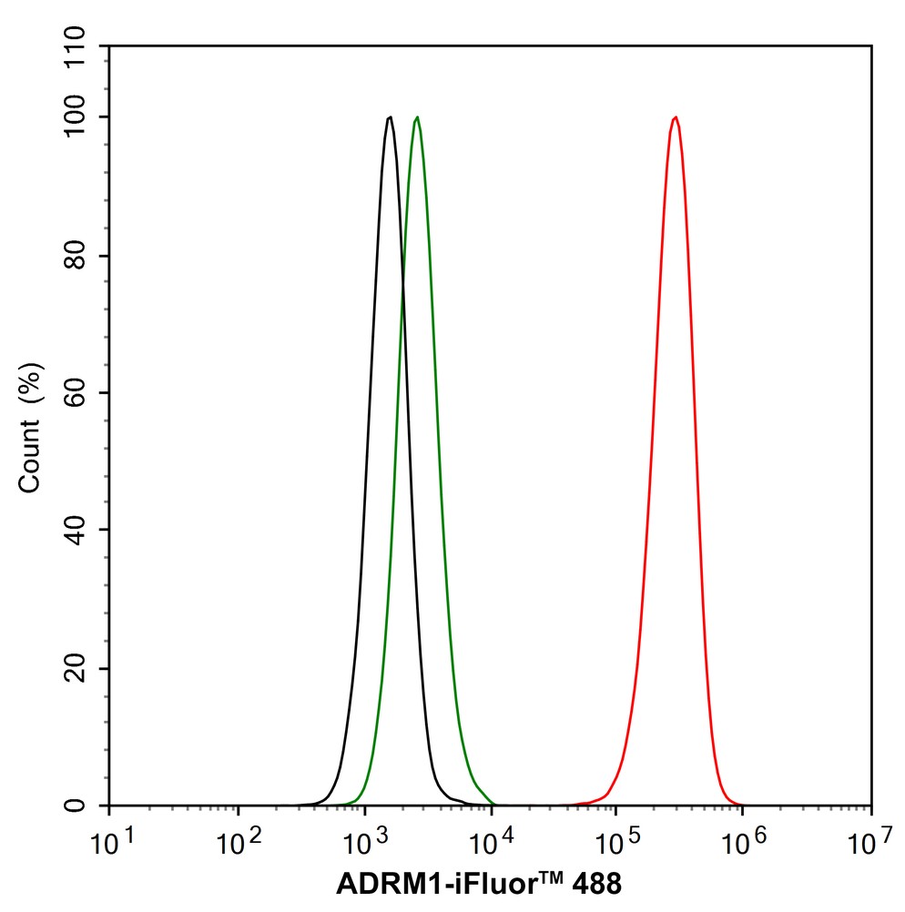
Flow cytometric analysis of NIH/3T3 cells labeling ADRM1. Cells were fixed and permeabilized. Then stained with the primary antibody (1/1,000) (red) compared with Rabbit IgG Isotype Control (green). After incubation of the primary antibody at +4℃ for an hour, the cells were stained with a 488 conjugate-Goat anti-Rabbit IgG Secondary antibody at 1/1,000 dilution for 30 minutes at +4℃. Unlabelled sample was used as a control (cells without incubation with primary antibody; black).IP
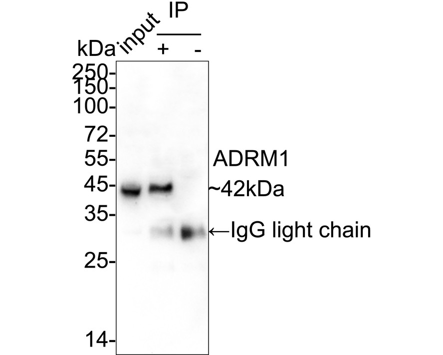
ADRM1 was immunoprecipitated from 0.2 mg HEK-293 cell lysate with Rabbit anti-ADRM1 antibody at 2 µg/10 µl beads. Western blot was performed from the immunoprecipitate using Rabbit anti-ADRM1 antibody at 1/1,000 dilution. Anti-Rabbit IgG for IP Nano-secondary antibody at 1/5,000 dilution was used for 1 hour at room temperature. Lane 1: HEK-293 cell lysate (input), Lane 2: Rabbit anti-ADRM1 antibody IP in HEK-293 cell lysate, Lane 3: Rabbit IgG instead of Rabbit anti-ADRM1 antibody in HEK-293 cell lysate, Blocking/Dilution buffer: 5% NFDM/TBST, Exposure time: 3 minutes.| Product Name | ADRM1 Recombinant Rabbit Monoclonal Antibody |
|---|---|
| Antibody Type | Primary Antibodies |
| Immunogen | Recombinant protein within human ADRM1 aa 258-407 / 407. |
| Clonality | Monoclonal |
|---|---|
| Isotype | IgG |
| Host Species | Rabbit |
| Tested Applications | FCICC/IFIPWB |
| WB:1:2000 ICC/IF:1:500 FC:1:1000 IP:1-2μg/sample |
|
| Species Reactivity | HumanMonkeyMouseRat |
| Concentration | 1mg/ml |
| Purification | Protein A |
| Gene Symbol | ADRM1 |
|---|---|
| Gene Synonyms | ARM1 ARM-1 GP110 PSMD16 |
| Gene Full Name | ADRM1 26S proteasome ubiquitin receptor |
| Gene Summary | This gene encodes a member of the adhesion regulating molecule 1 protein family. The encoded protein is a component of the proteasome where it acts as a ubiquitin receptor and recruits the deubiquitinating enzyme, ubiquitin carboxyl-terminal hydrolase L5. Increased levels of the encoded protein are associated with increased cell adhesion, which is likely an indirect effect of this intracellular protein. Dysregulation of this gene has been implicated in carcinogenesis. Alternative splicing results in multiple transcript variants. [provided by RefSeq, Jul 2013] |
| Alternative Names | 110 kDa cell membrane glycoprotein antibody adhesion regulating molecule 1 antibody Adhesion-regulating molecule 1 antibody ADRM 1 antibody Adrm1 antibody ADRM1_HUMAN antibody ARM 1 antibody ARM-1 antibody ARM1 antibody Gp110 antibody 110 kDa cell membrane glycoprotein antibody adhesion regulating molecule 1 antibody Adhesion-regulating molecule 1 antibody ADRM 1 antibody Adrm1 antibody ADRM1_HUMAN antibody ARM 1 antibody ARM-1 antibody ARM1 antibody Gp110 antibody hRpn13 antibody M(r) 110 000 surface antigen antibody Proteasomal ubiquitin receptor ADRM1 antibody proteasome regulatory particle non ATPase 13 antibody Proteasome regulatory particle non-ATPase 13 antibody Regulatory particle non ATPase 13 antibody Rpn13 antibody Rpn13 homolog antibody |
| Molecular Weight(MW) | 42 kDa |
| Cellular Localization | Cytoplasm, Nucleus. |
WB
Western blot analysis of ADRM1 on different lysates with Rabbit anti-ADRM1 antibody at 1/2,000 dilution. Lane 1: HEK-293 cell lysate (20 µg/Lane), Lane 2: HeLa cell lysate (20 µg/Lane), Lane 3: SK-MEL-28 cell lysate (20 µg/Lane), Lane 4: MCF7 cell lysate (20 µg/Lane), Lane 5: Ramos cell lysate (20 µg/Lane), Lane 6: Raji cell lysate (20 µg/Lane), Lane 7: K-562 cell lysate (20 µg/Lane), Lane 8: U-2 OS cell lysate (20 µg/Lane), Lane 9: SW620 cell lysate (20 µg/Lane), Lane 10: COS-1 cell lysate (20 µg/Lane), Lane 11: NIH/3T3 cell lysate (20 µg/Lane), Lane 12: RAW264.7 cell lysate (20 µg/Lane), Lane 13: PC-12 cell lysate (20 µg/Lane), Lane 14: C6 cell lysate (20 µg/Lane), Exposure time: 3 minutes; 4-20% SDS-PAGE gel. Proteins were transferred to a PVDF membrane and blocked with 5% NFDM/TBST for 1 hour at room temperature. The primary antibody at 1/2,000 dilution was used in 5% NFDM/TBST at 4℃ overnight. Goat Anti-Rabbit IgG - HRP Secondary Antibody at 1/50,000 dilution was used for 1 hour at room temperature.
ICC/IF
Immunocytochemistry analysis of SK-MEL-28 cells labeling ADRM1 with Rabbit anti-ADRM1 antibody at 1/500 dilution. Cells were fixed in 4% paraformaldehyde for 20 minutes at room temperature, permeabilized with 0.1% Triton X-100 in PBS for 5 minutes at room temperature, then blocked with 1% BSA in 10% negative goat serum for 1 hour at room temperature. Cells were then incubated with Rabbit anti-ADRM1 antibody at 1/500 dilution in 1% BSA in PBST overnight at 4 ℃. Goat Anti-Rabbit IgG H&L (488) was used as the secondary antibody at 1/1,000 dilution. PBS instead of the primary antibody was used as the secondary antibody only control. Nuclear DNA was labelled in blue with DAPI. Beta tubulin (red) was stained at 1/100 dilution overnight at +4℃. Goat Anti-Mouse IgG H&L (594) was used as the secondary antibody at 1/1,000 dilution.
FC
Flow cytometric analysis of NIH/3T3 cells labeling ADRM1. Cells were fixed and permeabilized. Then stained with the primary antibody (1/1,000) (red) compared with Rabbit IgG Isotype Control (green). After incubation of the primary antibody at +4℃ for an hour, the cells were stained with a 488 conjugate-Goat anti-Rabbit IgG Secondary antibody at 1/1,000 dilution for 30 minutes at +4℃. Unlabelled sample was used as a control (cells without incubation with primary antibody; black).
IP
ADRM1 was immunoprecipitated from 0.2 mg HEK-293 cell lysate with Rabbit anti-ADRM1 antibody at 2 µg/10 µl beads. Western blot was performed from the immunoprecipitate using Rabbit anti-ADRM1 antibody at 1/1,000 dilution. Anti-Rabbit IgG for IP Nano-secondary antibody at 1/5,000 dilution was used for 1 hour at room temperature. Lane 1: HEK-293 cell lysate (input), Lane 2: Rabbit anti-ADRM1 antibody IP in HEK-293 cell lysate, Lane 3: Rabbit IgG instead of Rabbit anti-ADRM1 antibody in HEK-293 cell lysate, Blocking/Dilution buffer: 5% NFDM/TBST, Exposure time: 3 minutes.| Application Notes | WB:1:2000 ICC/IF:1:500 FC:1:1000 IP:1-2μg/sample |
|---|
| Form | Liquid |
|---|---|
| Storage Instructions | Shipped at 4℃. Store at +4℃ short term (1-2 weeks). It is recommended to aliquot into single-use upon delivery. Store at -20℃ long term. |
| Storage Buffer | PBS (pH7.4), 0.1% BSA, 40% Glycerol. Preservative: 0.05% Sodium Azide. |


