WB
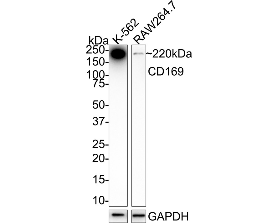
Western blot analysis of CD169 on different lysates with Rabbit anti-CD169 antibody at 1/2,000 dilution. Lane 1: K-562 cell lysate, Lane 2: RAW264.7 cell lysate, Lysates/proteins at 20 µg/Lane. Exposure time: 28 seconds; 4-20% SDS-PAGE gel. Proteins were transferred to a PVDF membrane and blocked with 5% NFDM/TBST for 1 hour at room temperature. The primary antibody at 1/2,000 dilution was used in 5% NFDM/TBST at 4℃ overnight. Goat Anti-Rabbit IgG - HRP Secondary Antibody at 1/50,000 dilution was used for 1 hour at room temperature.FC
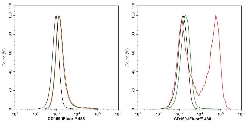
Flow cytometric analysis of THP-1 cells (left) and THP-1 cells treated with 50ng/mL PMA for 72 hours add 1μg/mL LPS for 24 hours (right) labeling CD169. Cells were washed twice with cold PBS and resuspend. Then stained with the primary antibody (0.5μg/mL) (red) compared with Rabbit IgG Isotype Control (green). After incubation of the primary antibody at +4℃ for an hour, the cells were stained with a 488 conjugate-Goat anti-Rabbit IgG Secondary antibody at 1/1,000 dilution for 30 minutes at +4℃. Unlabelled sample was used as a control (cells without incubation with primary antibody; black).| Product Name | CD169 Recombinant Rabbit Monoclonal Antibody |
|---|---|
| Antibody Type | Primary Antibodies |
| Immunogen | Recombinant protein within human CD169 aa 1-1,000 / 1,709. |
| Clonality | Monoclonal |
|---|---|
| Isotype | IgG |
| Host Species | Rabbit |
| Tested Applications | FCWB |
| WB:1:2000 FC:1:2000 |
|
| Species Reactivity | HumanMouse |
| Concentration | 1mg/ml |
| Purification | Protein A |
| Gene Symbol | SIGLEC1 |
|---|---|
| Gene Synonyms | SN CD169 SIGLEC-1 |
| Gene Full Name | sialic acid binding Ig like lectin 1 |
| Gene Summary | This gene encodes a member of the immunoglobulin superfamily. The encoded protein is a lectin-like adhesion molecule that binds glycoconjugate ligands on cell surfaces in a sialic acid-dependent manner. It is a type I transmembrane protein expressed only by a subpopulation of macrophages and is involved in mediating cell-cell interactions. The protein plays an important role in multiple human diseases and bacterial and viral infections has been shown to enhance SARS-CoV-2 infection. [provided by RefSeq, Dec 2021] |
| Alternative Names | CD169 antibody CD169 antigen antibody dJ1009E24.1 antibody DKFZp667F058 antibody FLJ00051 antibody FLJ00055 antibody FLJ00073 antibody FLJ00411 antibody FLJ32150 antibody Sialic acid binding Ig like lectin 1 antibody CD169 antibody CD169 antigen antibody dJ1009E24.1 antibody DKFZp667F058 antibody FLJ00051 antibody FLJ00055 antibody FLJ00073 antibody FLJ00411 antibody FLJ32150 antibody Sialic acid binding Ig like lectin 1 antibody sialic acid binding Ig-like lectin 1 sialoadhesin antibody Sialic acid binding immunoglobulin like lectin 1 antibody Sialic acid-binding Ig-like lectin 1 antibody Sialoadhesin antibody Siglec 1 antibody Siglec-1 antibody Siglec1 antibody SN antibody SN_HUMAN antibody |
| Molecular Weight(MW) | 183 kDa(Observed band size: 220 kDa) |
| Cellular Localization | Cell membrane, Secreted. |
WB
Western blot analysis of CD169 on different lysates with Rabbit anti-CD169 antibody at 1/2,000 dilution. Lane 1: K-562 cell lysate, Lane 2: RAW264.7 cell lysate, Lysates/proteins at 20 µg/Lane. Exposure time: 28 seconds; 4-20% SDS-PAGE gel. Proteins were transferred to a PVDF membrane and blocked with 5% NFDM/TBST for 1 hour at room temperature. The primary antibody at 1/2,000 dilution was used in 5% NFDM/TBST at 4℃ overnight. Goat Anti-Rabbit IgG - HRP Secondary Antibody at 1/50,000 dilution was used for 1 hour at room temperature.
FC
Flow cytometric analysis of THP-1 cells (left) and THP-1 cells treated with 50ng/mL PMA for 72 hours add 1μg/mL LPS for 24 hours (right) labeling CD169. Cells were washed twice with cold PBS and resuspend. Then stained with the primary antibody (0.5μg/mL) (red) compared with Rabbit IgG Isotype Control (green). After incubation of the primary antibody at +4℃ for an hour, the cells were stained with a 488 conjugate-Goat anti-Rabbit IgG Secondary antibody at 1/1,000 dilution for 30 minutes at +4℃. Unlabelled sample was used as a control (cells without incubation with primary antibody; black).| Application Notes | WB:1:2000 FC:1:2000 |
|---|
| Form | Liquid |
|---|---|
| Storage Instructions | Shipped at 4℃. Store at +4℃ short term (1-2 weeks). It is recommended to aliquot into single-use upon delivery. Store at -20℃ long term. |
| Storage Buffer | PBS (pH7.4), 0.1% BSA, 40% Glycerol. Preservative: 0.05% Sodium Azide. |


