WB
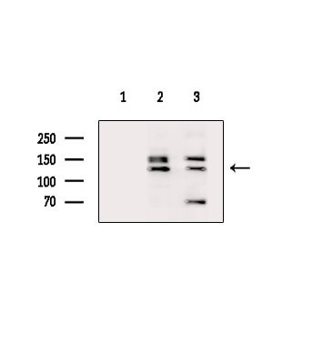
Western blot analysis of extracts from various samples, using RHG20 Antibody. Lane 1: 3t3-l1 cells(lps 4h treatment), blocked with antigen-specific peptides. Lane 2: 3t3-l1 cells(lps 4h treatment). Lane 3: Raw264.7 cells(h202 treatment).IHC
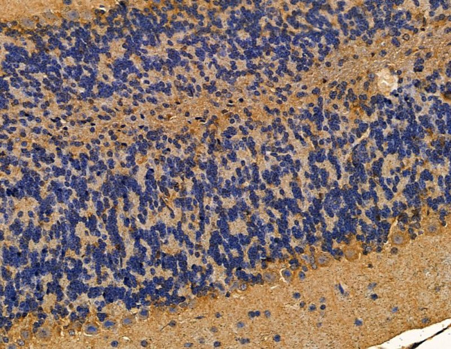
RHG20 Antibody at 1/100 staining mouse brain tissue by IHC-P. The sample was formaldehyde fixed and a heat mediated antigen retrieval step in citrate buffer was performed. The sample was then blocked and incubated with the primary antibody at 4°C overnight. An HRP conjugated anti-rabbit antibody was used as the secondary antibody.IHC
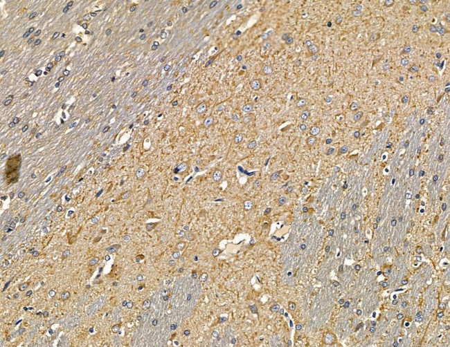
RHG20 Antibody at 1/100 at 1/100 staining rat brain tissue by IHC-P. The sample was formaldehyde fixed and a heat mediated antigen retrieval step in citrate buffer was performed. The sample was then blocked and incubated with the primary antibody at 4°C overnight. An HRP conjugated anti-rabbit antibody was used as the secondary antibody.| Product Name | Rabbit polyclonal antibody to RHG20 |
|---|---|
| Antibody Type | Primary Antibodies |
| Immunogen | A synthesized peptide derived from human RHG20, corresponding to a region within the internal amino acids. |
| Clonality | Polyclonal |
|---|---|
| Isotype | IgG |
| Host Species | Rabbit |
| Tested Applications | IHCWB |
| WB:1:1000-1:3000 IHC:1:50-1:200 |
|
| Species Reactivity | HumanMouseRat |
| Concentration | 1mg/ml |
| Purification | Affinity purified |
| Gene Symbol | ARHGAP20 |
|---|---|
| Gene Synonyms | RARHOGAP |
| Gene Full Name | Rho GTPase activating protein 20 |
| Gene Summary | The protein encoded by this gene is an activator of RHO-type GTPases, transducing a signal from RAP1 to RHO and impacting neurite outgrowth. [provided by RefSeq, Sep 2016] |
| Alternative Names | Arhgap20 KIAA1391 RARHOGAP RHG20_HUMAN Rho GTPase activating protein 20 Rho GTPase-activating protein 20 Rho type GTPase activating protein 20 Rho-type GTPase-activating protein 20 |
| Molecular Weight(MW) | 133kDa |
| Cellular Localization | cytosol. |
WB
Western blot analysis of extracts from various samples, using RHG20 Antibody. Lane 1: 3t3-l1 cells(lps 4h treatment), blocked with antigen-specific peptides. Lane 2: 3t3-l1 cells(lps 4h treatment). Lane 3: Raw264.7 cells(h202 treatment).
IHC
RHG20 Antibody at 1/100 staining mouse brain tissue by IHC-P. The sample was formaldehyde fixed and a heat mediated antigen retrieval step in citrate buffer was performed. The sample was then blocked and incubated with the primary antibody at 4°C overnight. An HRP conjugated anti-rabbit antibody was used as the secondary antibody.
IHC
RHG20 Antibody at 1/100 at 1/100 staining rat brain tissue by IHC-P. The sample was formaldehyde fixed and a heat mediated antigen retrieval step in citrate buffer was performed. The sample was then blocked and incubated with the primary antibody at 4°C overnight. An HRP conjugated anti-rabbit antibody was used as the secondary antibody.| Application Notes | WB:1:1000-1:3000 IHC:1:50-1:200 |
|---|
| Form | Liquid |
|---|---|
| Storage Instructions | Store at -20 °C. Stable for 12 months from date of receipt. |
| Storage Buffer | Rabbit IgG in phosphate buffered saline , pH 7.4, 150mM NaCl, 0.02% sodium azide and 50% glycerol. |


