WB
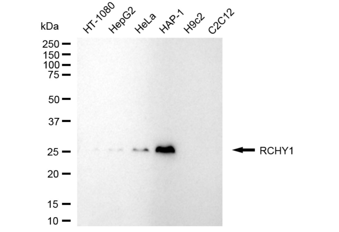
Western blotting analysis using RCHY1 antibody. Total cell lysates (30 μg) from various cell lines were loaded and separated by SDS-PAGE. The blot was incubated with RCHY1 antibody and HRP-conjugated goat anti-rabbit secondary antibody respectively.ICC/IF
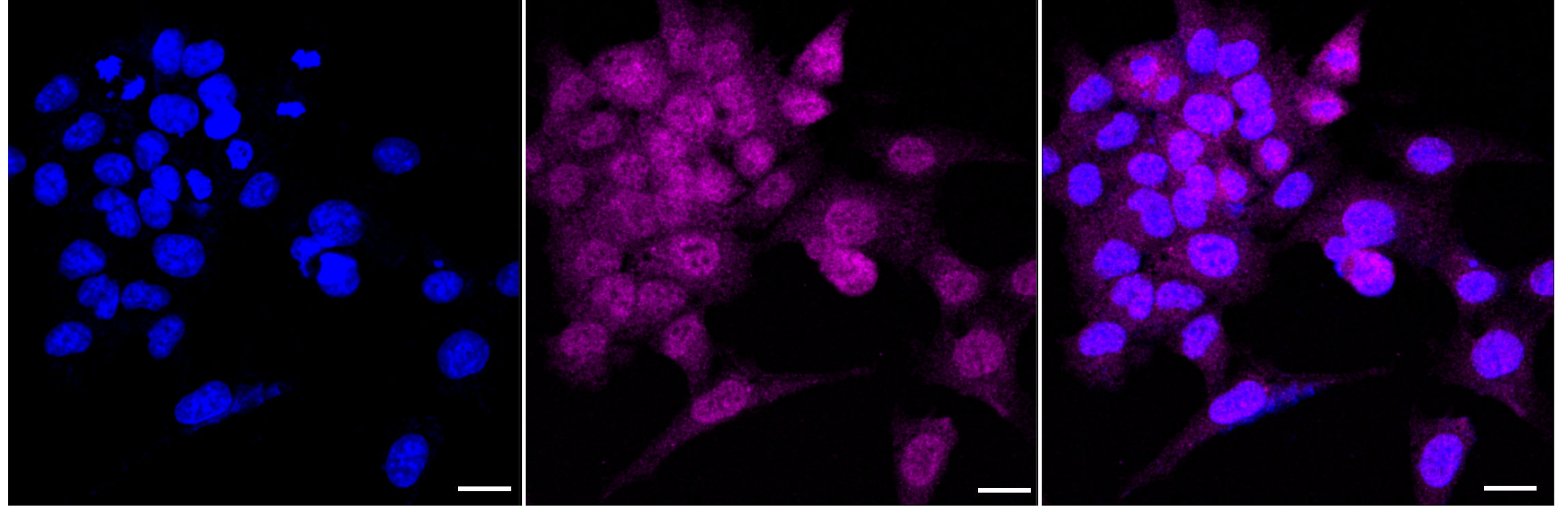
Immunocytochemical staining of HAP-1 cells with RCHY1 antibody. Nuclei were stained blue with DAPI; RCHY1 was stained magenta with Alexa Fluor® 647. Images were taken using Leica stellaris 5. Protein abundance based on laser Intensity and smart gain: Medium. Scale bar: 20 μm.FC
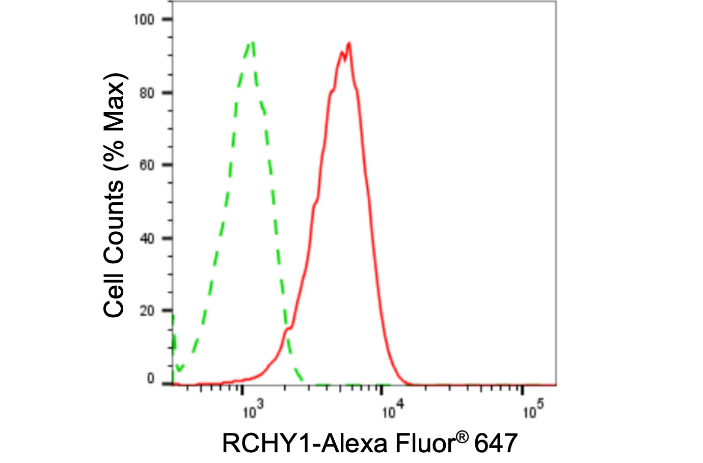
Flow cytometric analysis of RCHY1 expression in HAP-1 cells using RCHY1 antibody. Green, isotype control; red, RCHY1.| Product Name | RCHY1 Recombinant Monoclonal Antibody |
|---|---|
| Antibody Type | Primary Antibodies |
| Immunogen | Recombinant Human RCHY1 protein. |
| Clonality | Monoclonal |
|---|---|
| Isotype | IgG |
| Host Species | Rabbit |
| Tested Applications | FCICC/IFWB |
| WB:1:1000-1:5000 ICC/IF:1:100-1:1000 FC:1:200-1:2000 |
|
| Species Reactivity | Human |
| Concentration | 1mg/ml |
| Purification | Affinity purified |
| Gene Symbol | RCHY1 |
|---|---|
| Gene Synonyms | ZCHY ARNIP CHIMP PIRH2 RNF199 ZNF363 PRO1996 |
| Gene Full Name | ring finger and CHY zinc finger domain containing 1 |
| Gene Summary | The protein encoded by this gene has ubiquitin ligase activity. It mediates E3-dependent ubiquitination and proteasomal degradation of target proteins, including tumor protein 53, histone deacetylase 1, and cyclin-dependent kinase inhibitor 1B, thus regulating their levels and cell cycle progression. Alternatively spliced transcript variants encoding different isoforms have been described for this gene. [provided by RefSeq, Jun 2013] |
| Alternative Names | RCHY1 Ring Finger And CHY Zinc Finger Domain Containing 1 RNF199 ZNF363 CHIMP ARNIP PIRH2 PRO1996 ZCHY RING Finger And CHY Zinc Finger Domain-Containing Protein 1 Androgen-Receptor N-Terminal-Interacting Protein P53-Induced Protein With A RING-H2 Domain RING-Type E3 Ubiquitin Transferase RCHY1 E3 Ubiquitin-Protein Ligase Pirh2 RING Finger Protein 199 Zinc Finger Protein 363 Zinc Finger CHY-Type DKFZp586C1620 Ring Finger And CHY Zinc Finger Domain Containing 1 E3 Ubiquitin Protein Ligase Androgen Receptor N-Terminal-Interacting Protein CH-Rich Interacting Match With PLAG1 CH-Rich-Interacting Match With PLAG1 P53-Induced RING-H2 Protein EC 2.3.2.27 HPirh2 |
| Cellular Localization | Nucleus. Nucleus speckle. Cytoplasm. |
WB
Western blotting analysis using RCHY1 antibody. Total cell lysates (30 μg) from various cell lines were loaded and separated by SDS-PAGE. The blot was incubated with RCHY1 antibody and HRP-conjugated goat anti-rabbit secondary antibody respectively.
ICC/IF
Immunocytochemical staining of HAP-1 cells with RCHY1 antibody. Nuclei were stained blue with DAPI; RCHY1 was stained magenta with Alexa Fluor® 647. Images were taken using Leica stellaris 5. Protein abundance based on laser Intensity and smart gain: Medium. Scale bar: 20 μm.
FC
Flow cytometric analysis of RCHY1 expression in HAP-1 cells using RCHY1 antibody. Green, isotype control; red, RCHY1.| Application Notes | WB:1:1000-1:5000 ICC/IF:1:100-1:1000 FC:1:200-1:2000 |
|---|
| Form | Liquid |
|---|---|
| Storage Instructions | Upon receipt, store at -20°C or -80°C. Avoid repeated freeze. |
| Storage Buffer | 0.03% Proclin 300 Constituents: 50% Glycerol, 0.01M PBS, PH 7.4 |


