WB
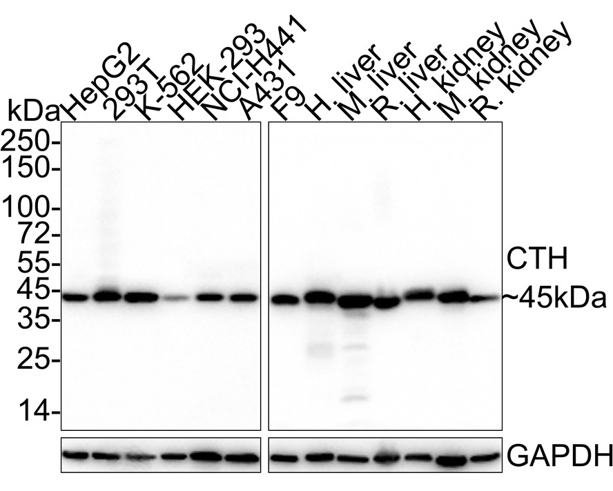
Western blot analysis of Cystathionase on different lysates with Rabbit anti-Cystathionase antibody at 1/2,000 dilution. Lane 1: HepG2 cell lysate (20 µg/Lane), Lane 2: 293T cell lysate (20 µg/Lane), Lane 3: K-562 cell lysate (20 µg/Lane), Lane 4: HEK-293 cell lysate (20 µg/Lane), Lane 5: NCI-H441 cell lysate (20 µg/Lane), Lane 6: A431 cell lysate (20 µg/Lane), Lane 7: F9 cell lysate (20 µg/Lane), Lane 8: Human liver tissue lysate (40 µg/Lane), Lane 9: Mouse liver tissue lysate (40 µg/Lane), Lane 10: Rat liver tissue lysate (40 µg/Lane), Lane 11: Human kidney tissue lysate (40 µg/Lane), Lane 12: Mouse kidney tissue lysate (40 µg/Lane), Lane 13: Rat kidney tissue lysate (40 µg/Lane), Exposure time: 8 seconds; 4-20% SDS-PAGE gel. Proteins were transferred to a PVDF membrane and blocked with 5% NFDM/TBST for 1 hour at room temperature. The primary antibody at 1/2,000 dilution was used in 5% NFDM/TBST at 4℃ overnight. Goat Anti-Rabbit IgG - HRP Secondary Antibody at 1/50,000 dilution was used for 1 hour at room temperature.IP
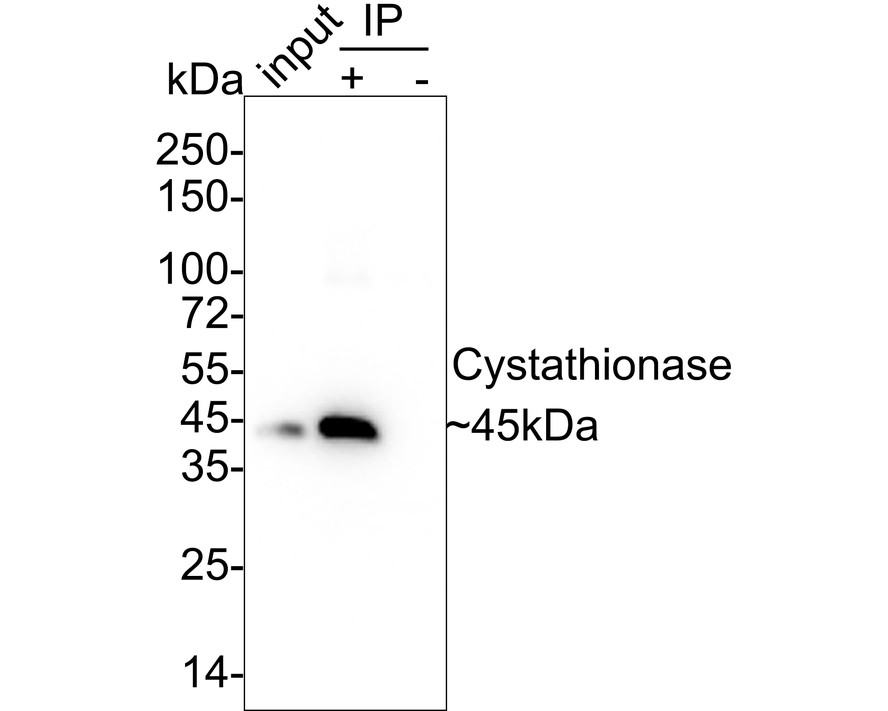
Cystathionase was immunoprecipitated in 0.2mg A431 cell lysate with Rabbit anti-Cystathionase antibody at 2 µg/10 µl beads. Western blot was performed from the immunoprecipitate using Rabbit anti-Cystathionase antibody at 1/1,000 dilution. Anti-Rabbit IgG for IP Nano-secondary antibody at 1/5,000 dilution was used for 1 hour at room temperature. Lane 1: A431 cell lysate (input), Lane 2: Rabbit anti-Cystathionase antibody IP in A431 cell lysate, Lane 3: Rabbit IgG instead of Rabbit anti-Cystathionase antibody in A431 cell lysate, Blocking/Dilution buffer: 5% NFDM/TBST, Exposure time: 24 seconds.| Product Name | Cystathionase Recombinant Rabbit Monoclonal Antibody |
|---|---|
| Antibody Type | Primary Antibodies |
| Immunogen | Recombinant protein within human Cystathionase aa 1-405 / 405. |
| Clonality | Monoclonal |
|---|---|
| Isotype | IgG |
| Host Species | Rabbit |
| Tested Applications | IPWB |
| WB:1:1000-1:10000 IP:1-2μg/sample |
|
| Species Reactivity | HumanMouseRat |
| Concentration | 1mg/ml |
| Purification | Protein A |
| Gene Symbol | CTH |
|---|---|
| Gene Synonyms | CGL CSE |
| Gene Full Name | cystathionine gamma-lyase |
| Gene Summary | This gene encodes a cytoplasmic enzyme in the trans-sulfuration pathway that converts cystathione derived from methionine into cysteine. Glutathione synthesis in the liver is dependent upon the availability of cysteine. Mutations in this gene cause cystathioninuria. Alternative splicing of this gene results in three transcript variants encoding different isoforms. [provided by RefSeq, Jun 2010] |
| Alternative Names | CGL_HUMAN antibody CTH antibody cystathionase (cystathionine gamma-lyase) antibody Cystathionine gamma lyase antibody Cystathionine gamma-lyase antibody Cysteine desulfhydrase antibody Gamma cystathionase antibody Gamma-cystathionase antibody Homoserine deaminase antibody Homoserine dehydratase antibody CGL_HUMAN antibody CTH antibody cystathionase (cystathionine gamma-lyase) antibody Cystathionine gamma lyase antibody Cystathionine gamma-lyase antibody Cysteine desulfhydrase antibody Gamma cystathionase antibody Gamma-cystathionase antibody Homoserine deaminase antibody Homoserine dehydratase antibody MGC9471 antibody |
| Molecular Weight(MW) | 45 kDa |
| Cellular Localization | Cytoplasm. |
WB
Western blot analysis of Cystathionase on different lysates with Rabbit anti-Cystathionase antibody at 1/2,000 dilution. Lane 1: HepG2 cell lysate (20 µg/Lane), Lane 2: 293T cell lysate (20 µg/Lane), Lane 3: K-562 cell lysate (20 µg/Lane), Lane 4: HEK-293 cell lysate (20 µg/Lane), Lane 5: NCI-H441 cell lysate (20 µg/Lane), Lane 6: A431 cell lysate (20 µg/Lane), Lane 7: F9 cell lysate (20 µg/Lane), Lane 8: Human liver tissue lysate (40 µg/Lane), Lane 9: Mouse liver tissue lysate (40 µg/Lane), Lane 10: Rat liver tissue lysate (40 µg/Lane), Lane 11: Human kidney tissue lysate (40 µg/Lane), Lane 12: Mouse kidney tissue lysate (40 µg/Lane), Lane 13: Rat kidney tissue lysate (40 µg/Lane), Exposure time: 8 seconds; 4-20% SDS-PAGE gel. Proteins were transferred to a PVDF membrane and blocked with 5% NFDM/TBST for 1 hour at room temperature. The primary antibody at 1/2,000 dilution was used in 5% NFDM/TBST at 4℃ overnight. Goat Anti-Rabbit IgG - HRP Secondary Antibody at 1/50,000 dilution was used for 1 hour at room temperature.
IP
Cystathionase was immunoprecipitated in 0.2mg A431 cell lysate with Rabbit anti-Cystathionase antibody at 2 µg/10 µl beads. Western blot was performed from the immunoprecipitate using Rabbit anti-Cystathionase antibody at 1/1,000 dilution. Anti-Rabbit IgG for IP Nano-secondary antibody at 1/5,000 dilution was used for 1 hour at room temperature. Lane 1: A431 cell lysate (input), Lane 2: Rabbit anti-Cystathionase antibody IP in A431 cell lysate, Lane 3: Rabbit IgG instead of Rabbit anti-Cystathionase antibody in A431 cell lysate, Blocking/Dilution buffer: 5% NFDM/TBST, Exposure time: 24 seconds.| Application Notes | WB:1:1000-1:10000 IP:1-2μg/sample |
|---|
| Form | Liquid |
|---|---|
| Storage Instructions | Shipped at 4℃. Store at +4℃ short term (1-2 weeks). It is recommended to aliquot into single-use upon delivery. Store at -20℃ long term. |
| Storage Buffer | PBS (pH7.4), 0.1% BSA, 40% Glycerol. Preservative: 0.05% Sodium Azide. |


