WB
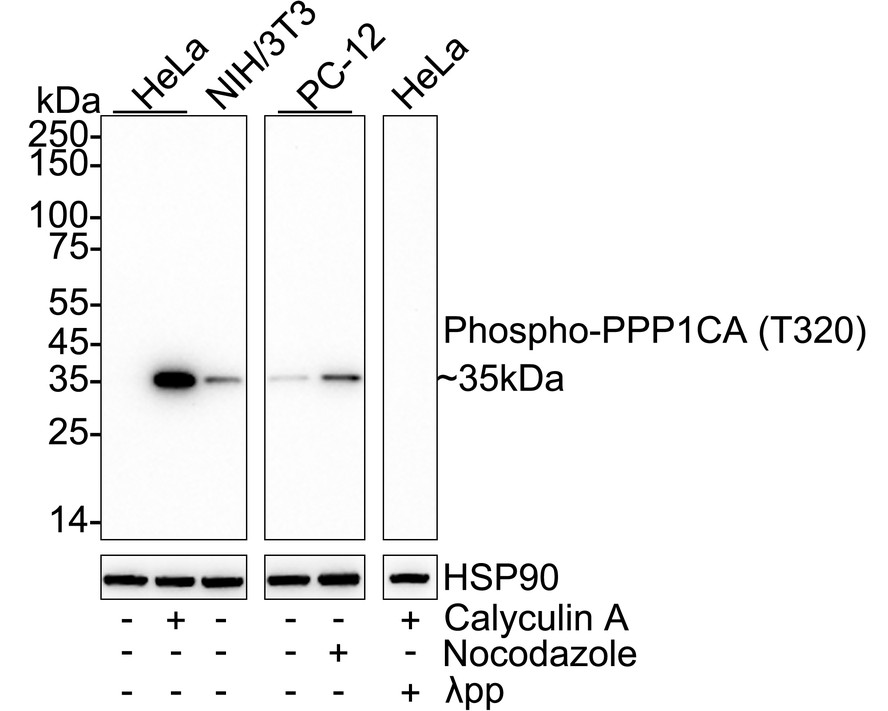
Western blot analysis of Phospho-PPP1CA (T320) on different lysates with Rabbit anti-Phospho-PPP1CA (T320) antibody at 1/2,000 dilution.Lysates/proteins at 20 µg/Lane.Exposure time: 3 minutes; 4-20% SDS-PAGE gel. Proteins were transferred to a PVDF membrane and blocked with 5% NFDM/TBST for 1 hour at room temperature. The primary antibody at 1/2,000 dilution was used in 5% NFDM/TBST at 4℃ overnight. Goat Anti-Rabbit IgG - HRP Secondary Antibody at 1/50,000 dilution was used for 1 hour at room temperature.IHC
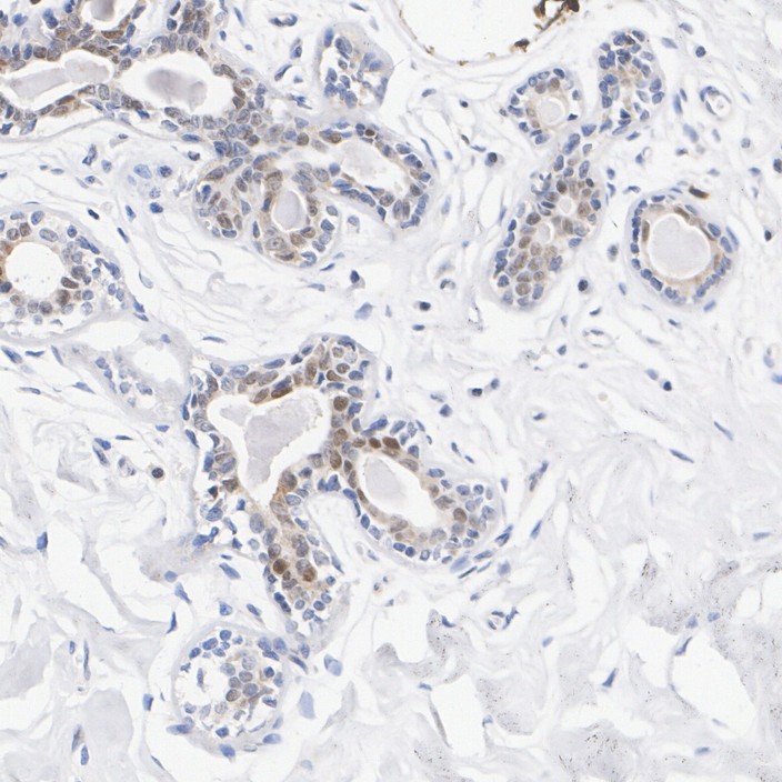
Immunohistochemical analysis of paraffin-embedded human breast cancer tissue with Rabbit anti-Phospho-PPP1CA (T320) antibody at 1/20,000 dilution.The section was pre-treated using heat mediated antigen retrieval with Tris-EDTA buffer (pH 9.0) for 20 minutes. The tissues were blocked in 1% BSA for 20 minutes at room temperature, washed with ddH2O and PBS, and then probed with the primary antibody at 1/20,000 dilution for 1 hour at room temperature. The detection was performed using an HRP conjugated compact polymer system. DAB was used as the chromogen. Tissues were counterstained with hematoxylin and mounted with DPX.IHC
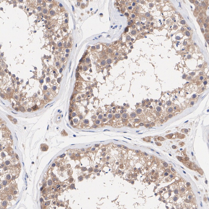
Immunohistochemical analysis of paraffin-embedded human testis tissue with Rabbit anti-Phospho-PPP1CA (T320) antibody at 1/20,000 dilution. The section was pre-treated using heat mediated antigen retrieval with Tris-EDTA buffer (pH 9.0) for 20 minutes. The tissues were blocked in 1% BSA for 20 minutes at room temperature, washed with ddH2O and PBS, and then probed with the primary antibody at 1/20,000 dilution for 1 hour at room temperature. The detection was performed using an HRP conjugated compact polymer system. DAB was used as the chromogen. Tissues were counterstained with hematoxylin and mounted with DPX.IHC
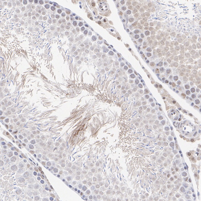
Immunohistochemical analysis of paraffin-embedded rat testis tissue with Rabbit anti-Phospho-PPP1CA (T320) antibody at 1/20,000 dilution. The section was pre-treated using heat mediated antigen retrieval with Tris-EDTA buffer (pH 9.0) for 20 minutes. The tissues were blocked in 1% BSA for 20 minutes at room temperature, washed with ddH2O and PBS, and then probed with the primary antibody at 1/20,000 dilution for 1 hour at room temperature. The detection was performed using an HRP conjugated compact polymer system. DAB was used as the chromogen. Tissues were counterstained with hematoxylin and mounted with DPX.IP
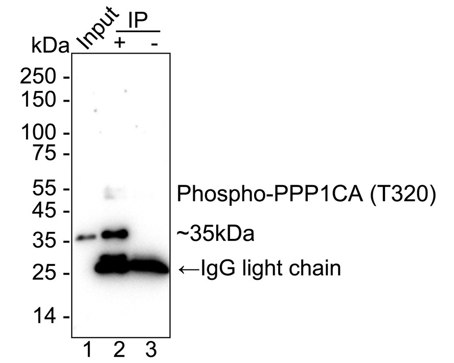
Phospho-PPP1CA (T320) was immunoprecipitated from 0.2 mg HeLa treated with 50mM Calyculin A for 3 hours cell lysate with Rabbit anti-Phospho-PPP1CA at 2 µg/10 µl beads. Western blot was performed from the immunoprecipitate using Rabbit anti-Phospho-PPP1CA at 1/2,000 dilution. Mouse Anti-Rabbit IgG kappa light chain secondary antibody at 1/5,000 dilution was used for 1 hour at room temperature. Blocking/Dilution buffer: 5% NFDM/TBST Exposure time: 1 minute 23 seconds;| Product Name | Phospho-PPP1CA (T320) Recombinant Rabbit Monoclonal Antibody |
|---|---|
| Antibody Type | Primary Antibodies |
| Immunogen | Synthetic phospho-peptide corresponding to residues surrounding Thr320 of Human PPP1CA. |
| Modification | p-T320 |
| Clonality | Monoclonal |
|---|---|
| Isotype | IgG |
| Host Species | Rabbit |
| Tested Applications | IHCIPWB |
| WB:1:2000 IHC:1:20000 IP:1-2μg/sample |
|
| Species Reactivity | HumanMouseRat |
| Concentration | 1mg/ml |
| Purification | Protein A |
| Gene Symbol | PPP1CA |
|---|---|
| Gene Synonyms | PP1A PP-1A PPP1A PP1alpha |
| Gene Full Name | protein phosphatase 1 catalytic subunit alpha |
| Gene Summary | The protein encoded by this gene is one of the three catalytic subunits of protein phosphatase 1 (PP1). This broadly expressed gene encodes the alpha subunit of the PP1 complex that associates with over 200 regulatory proteins to form holoenzymes which dephosphorylate their biological targets with high specificity. PP1 is a serine/threonine specific protein phosphatase known to be involved in the regulation of a variety of cellular processes, such as cell division, glycogen metabolism, muscle contractility, protein synthesis, and HIV-1 viral transcription. Increased PP1 activity has been observed in the end stage of heart failure. Studies suggest that PP1 is an important regulator of cardiac function and that PP1 deregulation is implicated in diabetes and multiple types of cancer. Three alternatively spliced transcript variants encoding different isoforms have been found for this gene. [provided by RefSeq, Jul 2020] |
| Alternative Names | Alpha isoform serine threonine protein phosphatase PP1alpha 1 catalytic subunit antibody Catalytic subunit antibody EC 3.1.3.16 antibody MGC15877 antibody MGC1674 antibody PP 1A antibody PP-1A antibody PP1A antibody PP1A_HUMAN antibody PP1alpha antibody Alpha isoform serine threonine protein phosphatase PP1alpha 1 catalytic subunit antibody Catalytic subunit antibody EC 3.1.3.16 antibody MGC15877 antibody MGC1674 antibody PP 1A antibody PP-1A antibody PP1A antibody PP1A_HUMAN antibody PP1alpha antibody PP2C ALPHA antibody PP2CA antibody Ppp1ca antibody Protein Phosphatase 2C Alpha Isoform antibody Serine threonine protein phosphatase PP1 alpha catalytic subunit antibody Serine threonine protein phosphatase PP1 alpha catalytic subunit protein phosphatase 1 antibody Serine/threonine-protein phosphatase PP1-alpha catalytic subunit antibody |
| Molecular Weight(MW) | 38 kDa(Observed band size: 35 kDa) |
| Cellular Localization | Cytoplasm, Nucleus, nucleoplasm, nucleolus. |
WB
Western blot analysis of Phospho-PPP1CA (T320) on different lysates with Rabbit anti-Phospho-PPP1CA (T320) antibody at 1/2,000 dilution.Lysates/proteins at 20 µg/Lane.Exposure time: 3 minutes; 4-20% SDS-PAGE gel. Proteins were transferred to a PVDF membrane and blocked with 5% NFDM/TBST for 1 hour at room temperature. The primary antibody at 1/2,000 dilution was used in 5% NFDM/TBST at 4℃ overnight. Goat Anti-Rabbit IgG - HRP Secondary Antibody at 1/50,000 dilution was used for 1 hour at room temperature.
IHC
Immunohistochemical analysis of paraffin-embedded human breast cancer tissue with Rabbit anti-Phospho-PPP1CA (T320) antibody at 1/20,000 dilution.The section was pre-treated using heat mediated antigen retrieval with Tris-EDTA buffer (pH 9.0) for 20 minutes. The tissues were blocked in 1% BSA for 20 minutes at room temperature, washed with ddH2O and PBS, and then probed with the primary antibody at 1/20,000 dilution for 1 hour at room temperature. The detection was performed using an HRP conjugated compact polymer system. DAB was used as the chromogen. Tissues were counterstained with hematoxylin and mounted with DPX.
IHC
Immunohistochemical analysis of paraffin-embedded human testis tissue with Rabbit anti-Phospho-PPP1CA (T320) antibody at 1/20,000 dilution. The section was pre-treated using heat mediated antigen retrieval with Tris-EDTA buffer (pH 9.0) for 20 minutes. The tissues were blocked in 1% BSA for 20 minutes at room temperature, washed with ddH2O and PBS, and then probed with the primary antibody at 1/20,000 dilution for 1 hour at room temperature. The detection was performed using an HRP conjugated compact polymer system. DAB was used as the chromogen. Tissues were counterstained with hematoxylin and mounted with DPX.
IHC
Immunohistochemical analysis of paraffin-embedded rat testis tissue with Rabbit anti-Phospho-PPP1CA (T320) antibody at 1/20,000 dilution. The section was pre-treated using heat mediated antigen retrieval with Tris-EDTA buffer (pH 9.0) for 20 minutes. The tissues were blocked in 1% BSA for 20 minutes at room temperature, washed with ddH2O and PBS, and then probed with the primary antibody at 1/20,000 dilution for 1 hour at room temperature. The detection was performed using an HRP conjugated compact polymer system. DAB was used as the chromogen. Tissues were counterstained with hematoxylin and mounted with DPX.
IP
Phospho-PPP1CA (T320) was immunoprecipitated from 0.2 mg HeLa treated with 50mM Calyculin A for 3 hours cell lysate with Rabbit anti-Phospho-PPP1CA at 2 µg/10 µl beads. Western blot was performed from the immunoprecipitate using Rabbit anti-Phospho-PPP1CA at 1/2,000 dilution. Mouse Anti-Rabbit IgG kappa light chain secondary antibody at 1/5,000 dilution was used for 1 hour at room temperature. Blocking/Dilution buffer: 5% NFDM/TBST Exposure time: 1 minute 23 seconds;| Application Notes | WB:1:2000 IHC:1:20000 IP:1-2μg/sample |
|---|
| Form | Liquid |
|---|---|
| Storage Instructions | Shipped at 4℃. Store at +4℃ short term (1-2 weeks). It is recommended to aliquot into single-use upon delivery. Store at -20℃ long term. |
| Storage Buffer | PBS (pH7.4), 0.1% BSA, 40% Glycerol. Preservative: 0.05% Sodium Azide. |


