WB
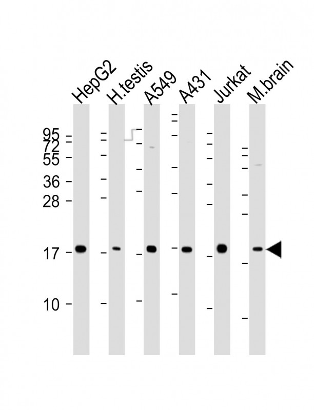
All lanes : Anti-EEF1E1 Antibody at 1:2000-1:4000 dilution; ; Lysates/proteins at 20 μg per lane. Secondary Goat Anti-mouse IgG, (H+L), Peroxidase conjugated at 1/10000 dilution.Blocking/Dilution buffer: 5% NFDM/TBST.ICC/IF
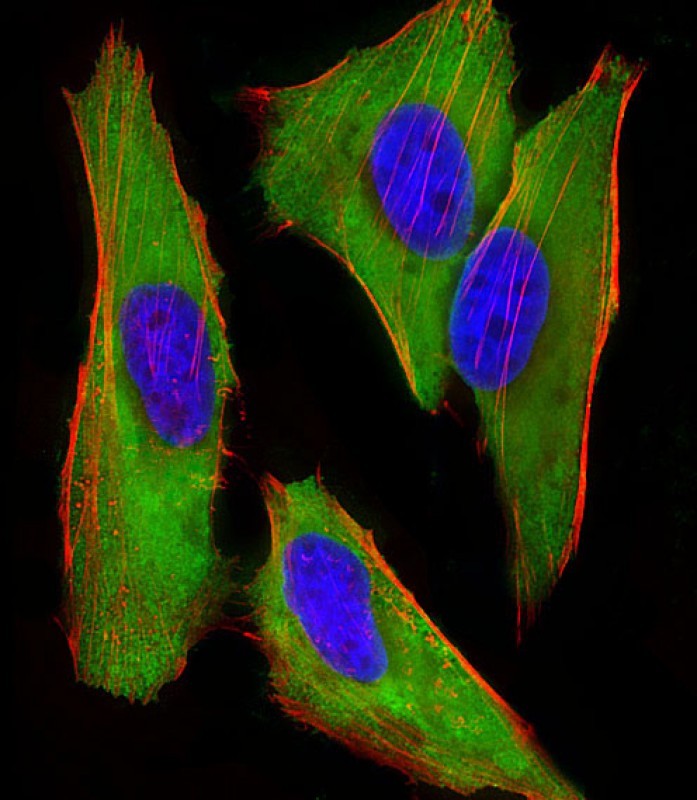
Immunofluorescent analysis of 4% paraformaldehyde-fixed, 0.1% Triton X-100 permeabilized HeLa (human cervical epithelial adenocarcinoma cell line) cells labeling EEF1E1 with Anti-EEF1E1 Antibody at 1/25 dilution, followed by Dylight® 488-conjugated goat anti-mouse IgG secondary antibody at 1/200 dilution (green). Immunofluorescence image showing cytoplasm staining on HeLa cell line. Cytoplasmic actin is detected with Dylight® 554 Phalloidin at 1/100 dilution (red).The nuclear counter stain is DAPI (blue).FC
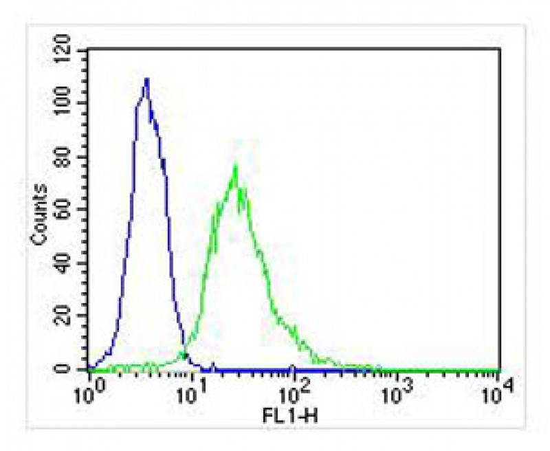
Overlay histogram showing Hela cells stained with Anti-EEF1E1 Antibody (green line). The cells were fixed with 2% paraformaldehyde (10 min) and then permeabilized with 90% methanol for 10 min. The cells were then icubated in 2% bovine serum albumin to block non-specific protein-protein interactions followed by the antibody (1:25 dilution) for 60 min at 37ºC. The secondary antibody used was Goat-Anti-Mouse IgG, DyLight® 488 Conjugated Highly Cross-Adsorbed) at 1/400 dilution for 40 min at 37ºC. Isotype control antibody (blue line) was mouse IgG2b (1μg/1x10^6 cells) used under the same conditions. Acquisition of >10, 000 events was performed.| Product Name | Mouse Monoclonal Antibody to EEF1E1 |
|---|---|
| Antibody Type | Primary Antibodies |
| Immunogen | This EEF1E1 antibody is generated from a mouse immunized with a recombinant protein of human EEF1E1. |
| Clonality | Monoclonal |
|---|---|
| Isotype | IgG2b |
| Host Species | Mouse |
| Tested Applications | FCICC/IFWB |
| WB:1:2000-1:4000 ICC/IF:1:25 FC:1:25 |
|
| Species Reactivity | HumanMouse |
| Concentration | 1mg/ml |
| Purification | Affinity purified |
| Gene Symbol | EEF1E1 |
|---|---|
| Gene Synonyms | P18 AIMP3 |
| Gene Full Name | eukaryotic translation elongation factor 1 epsilon 1 |
| Gene Summary | This gene encodes a multifunctional protein that localizes to both the cytoplasm and nucleus. In the cytoplasm, the encoded protein is an auxiliary component of the macromolecular aminoacyl-tRNA synthase complex. However, its mouse homolog has been shown to translocate to the nucleus in response to DNA damage, and it plays a positive role in ATM/ATR-mediated p53 activation. Alternative splicing results in multiple transcript variants. Read-through transcription also exists between this gene and the neighboring downstream MUTED (muted homolog) gene. An EEF1E1-related pseudogene has been identified on chromosome 2. [provided by RefSeq, Dec 2010] |
| Alternative Names | Eukaryotic translation elongation factor 1 epsilon-1 Aminoacyl tRNA synthetase complex-interacting multifunctional protein 3 Elongation factor p18 Multisynthase complex auxiliary component p18 EEF1E1 AIMP3 P18 |
| Molecular Weight(MW) | 20 kDa |
| Cellular Localization | Cytoplasm,Nucleus. |
WB
All lanes : Anti-EEF1E1 Antibody at 1:2000-1:4000 dilution; ; Lysates/proteins at 20 μg per lane. Secondary Goat Anti-mouse IgG, (H+L), Peroxidase conjugated at 1/10000 dilution.Blocking/Dilution buffer: 5% NFDM/TBST.
ICC/IF
Immunofluorescent analysis of 4% paraformaldehyde-fixed, 0.1% Triton X-100 permeabilized HeLa (human cervical epithelial adenocarcinoma cell line) cells labeling EEF1E1 with Anti-EEF1E1 Antibody at 1/25 dilution, followed by Dylight® 488-conjugated goat anti-mouse IgG secondary antibody at 1/200 dilution (green). Immunofluorescence image showing cytoplasm staining on HeLa cell line. Cytoplasmic actin is detected with Dylight® 554 Phalloidin at 1/100 dilution (red).The nuclear counter stain is DAPI (blue).
FC
Overlay histogram showing Hela cells stained with Anti-EEF1E1 Antibody (green line). The cells were fixed with 2% paraformaldehyde (10 min) and then permeabilized with 90% methanol for 10 min. The cells were then icubated in 2% bovine serum albumin to block non-specific protein-protein interactions followed by the antibody (1:25 dilution) for 60 min at 37ºC. The secondary antibody used was Goat-Anti-Mouse IgG, DyLight® 488 Conjugated Highly Cross-Adsorbed) at 1/400 dilution for 40 min at 37ºC. Isotype control antibody (blue line) was mouse IgG2b (1μg/1x10^6 cells) used under the same conditions. Acquisition of >10, 000 events was performed.| Application Notes | WB:1:2000-1:4000 ICC/IF:1:25 FC:1:25 |
|---|
| Form | Liquid |
|---|---|
| Storage Instructions | Store at 4°C short term. Aliquot and store at -20°C long term. Avoid freeze/thaw cycles. |
| Storage Buffer | Purified antibody in PBS with 0.05% sodium azide. |


