WB
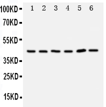
Western blot analysis of TRAM1 using anti-TRAM1 antibody. The sample well of each lane was loaded with 30 ug of sample under reducing conditions. Lane 1: rat brain tissue lysates, Lane 2: rat kidney tissue lysates, Lane 3: human 293T whole cell tissue lysates, Lane 4: human Raji whole cell lysates, Lane 5: human Jurkat whole cell lysates, Lane 6: human Hela whole cell lysates. After electrophoresis, proteins were transferred to a membrane. Then the membrane was incubated with rabbit anti-TRAM1 antigen affinity purified polyclonal antibody at a dilution of 1:1000 and probed with a goat anti-rabbit IgG-HRP secondary antibody. The signal is developed using ECL Plus Western Blotting Substrate.IHC
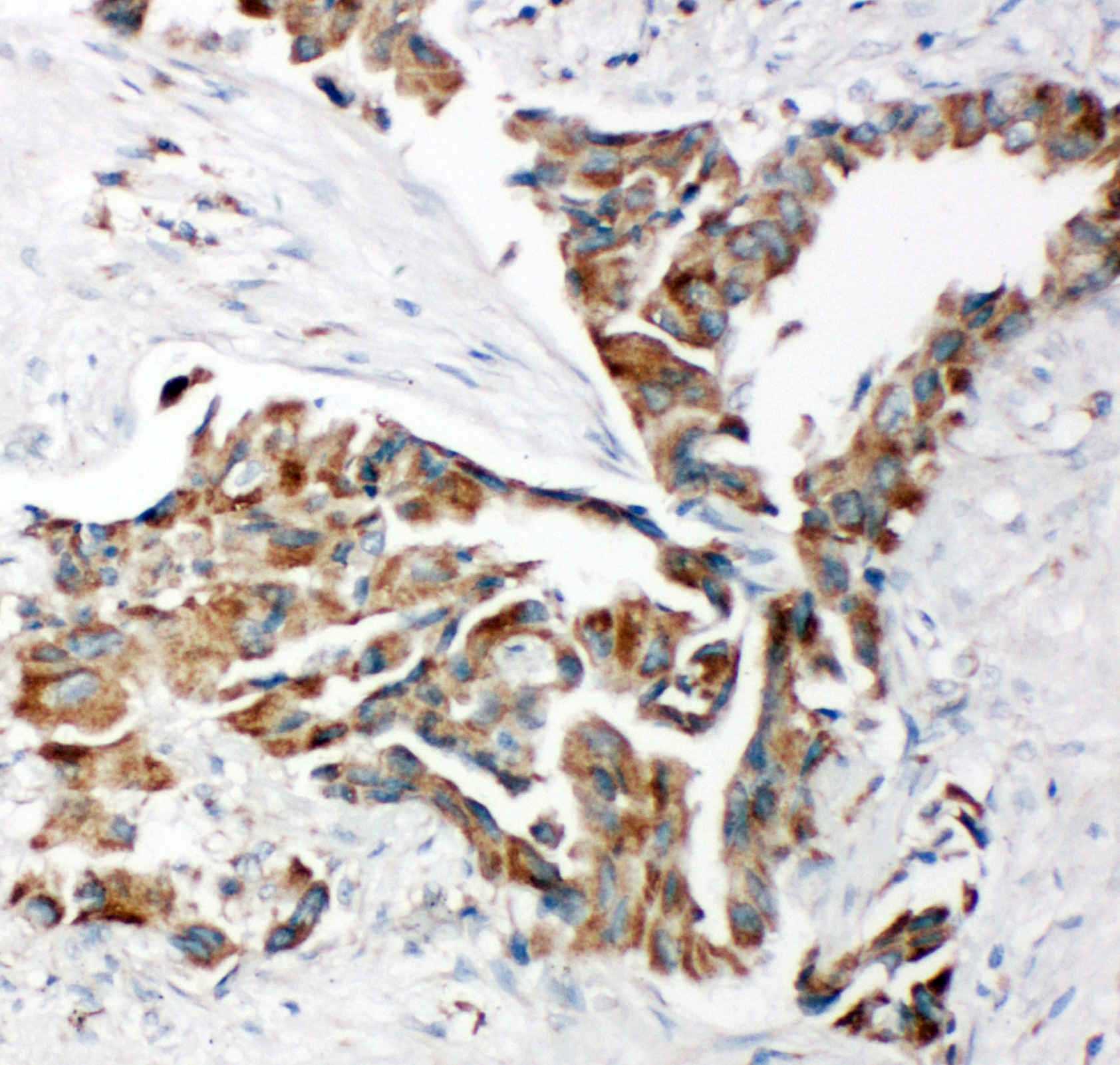
IHC analysis of TRAM1 using anti-TRAM1 antibody . TRAM1 was detected in a paraffin-embedded section of human lung cancer tissue. The tissue section was incubated with rabbit anti-TRAM1 Antibody at a dilution of 1:200 and developed using HRP Conjugated Rabbit IgG Super Vision Assay Kit with DAB as the chromogen.IHC
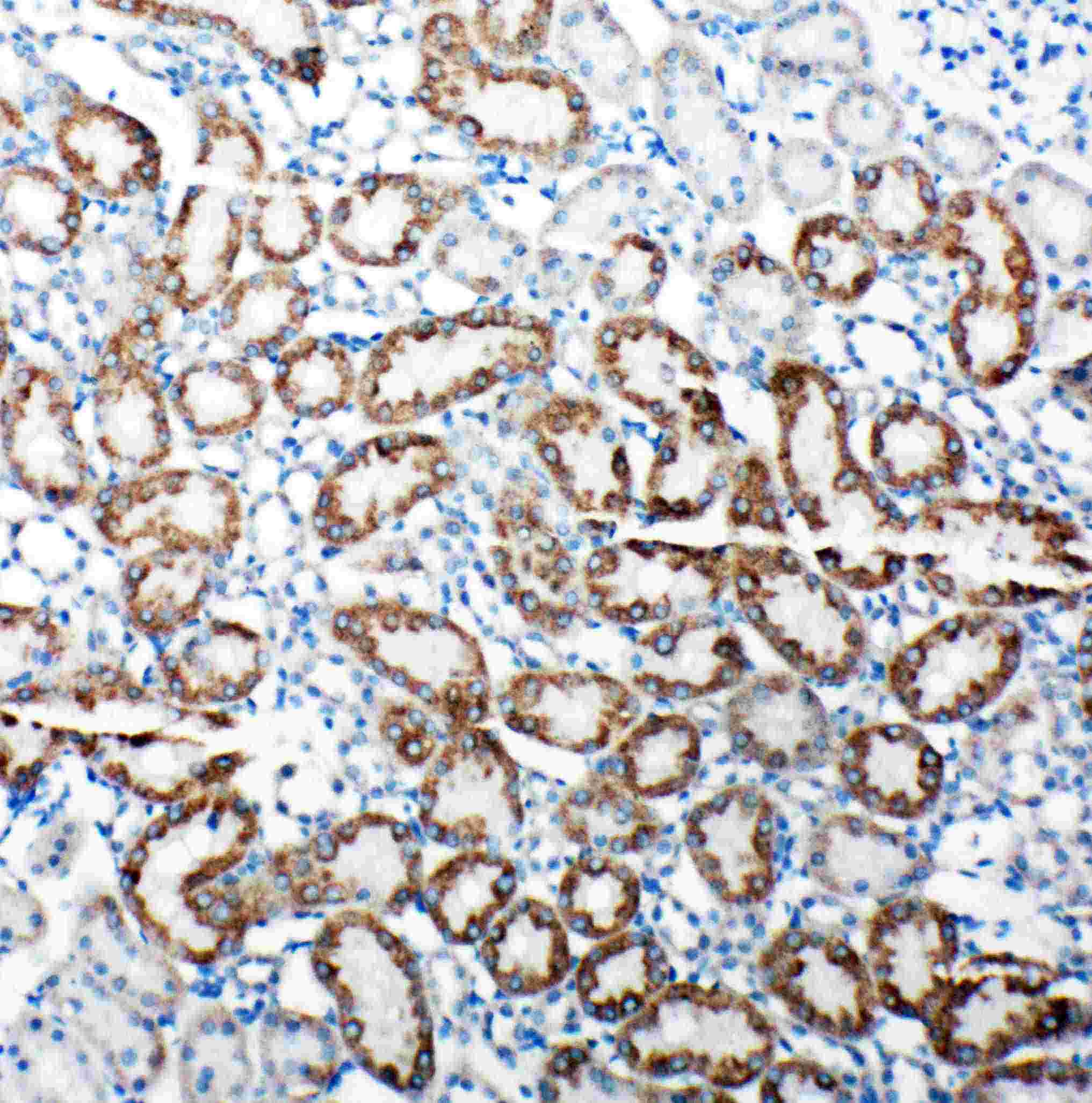
IHC analysis of TRAM1 using anti-TRAM1 antibody. TRAM1 was detected in a paraffin-embedded section of rat kidney tissue. The tissue section was incubated with rabbit anti-TRAM1 Antibody at a dilution of 1:200 and developed using HRP Conjugated Rabbit IgG Super Vision Assay Kit with DAB (Catalog # AR1027) as the chromogen.IHC
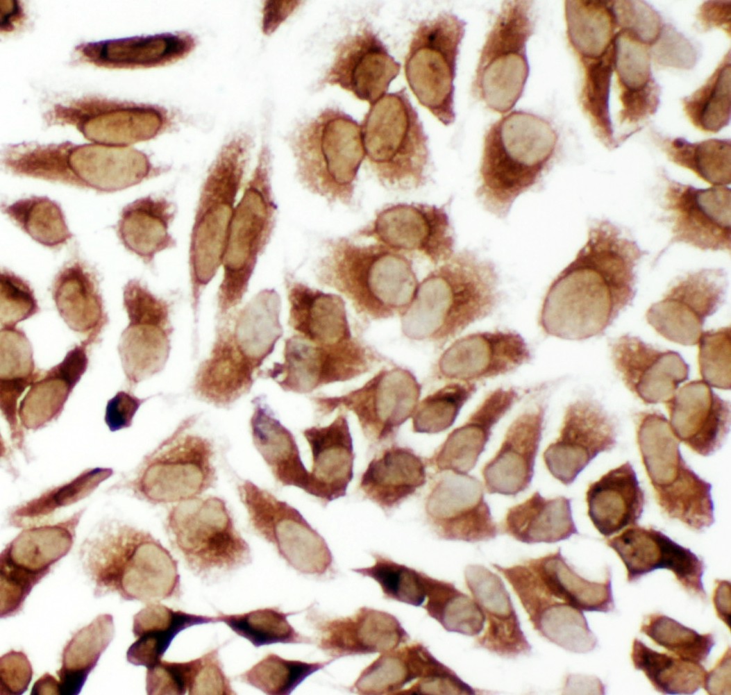
ICC analysis of TRAM1 using anti- TRAM1 antibody. TRAM1 was detected in an immunocytochemical section of Hela cells. The section was incubated with rabbit anti-TRAM1 Antibody at a dilution of 1:100 and developed using HRP Conjugated Rabbit IgG Super Vision Assay Kit with DAB as the chromogen.| Product Name | Rabbit polyclonal antibody to TRAM1 |
|---|---|
| Antibody Type | Primary Antibodies |
| Immunogen | A synthetic peptide corresponding to a sequence at the C-terminus of human TRAM1, identical to the related rat and mouse sequences. |
| Clonality | Polyclonal |
|---|---|
| Isotype | IgG |
| Host Species | Rabbit |
| Tested Applications | IHCWB |
| WB:1:500-1:2000 IHC:1:50-1:400 |
|
| Species Reactivity | HumanMouseRat |
| Concentration | 0.5mg/ml |
| Purification | Affinity purified |
| Gene Symbol | TRAM1 |
|---|---|
| Gene Synonyms | TRAM PNAS8 TRAMP |
| Gene Full Name | translocation associated membrane protein 1 |
| Gene Summary | This gene encodes a multi-pass membrane protein that is part of the mammalian endoplasmic reticulum. The encoded protein influences glycosylation and facilitates the translocation of secretory proteins across the endoplasmic reticulum membrane by regulating which domains of the nascent polypeptide chain are visible to the cytosol during a translocational pause. [provided by RefSeq, Oct 2009] |
| Alternative Names | PNAS 8 PRO1292 TRAM TRAM1 TRAMP |
| Molecular Weight(MW) | 43 kDa(Observed band size: 45,60 kDa) |
| Cellular Localization | Endoplasmic reticulum,Membrane |
WB
Western blot analysis of TRAM1 using anti-TRAM1 antibody. The sample well of each lane was loaded with 30 ug of sample under reducing conditions. Lane 1: rat brain tissue lysates, Lane 2: rat kidney tissue lysates, Lane 3: human 293T whole cell tissue lysates, Lane 4: human Raji whole cell lysates, Lane 5: human Jurkat whole cell lysates, Lane 6: human Hela whole cell lysates. After electrophoresis, proteins were transferred to a membrane. Then the membrane was incubated with rabbit anti-TRAM1 antigen affinity purified polyclonal antibody at a dilution of 1:1000 and probed with a goat anti-rabbit IgG-HRP secondary antibody. The signal is developed using ECL Plus Western Blotting Substrate.
IHC
IHC analysis of TRAM1 using anti-TRAM1 antibody . TRAM1 was detected in a paraffin-embedded section of human lung cancer tissue. The tissue section was incubated with rabbit anti-TRAM1 Antibody at a dilution of 1:200 and developed using HRP Conjugated Rabbit IgG Super Vision Assay Kit with DAB as the chromogen.
IHC
IHC analysis of TRAM1 using anti-TRAM1 antibody. TRAM1 was detected in a paraffin-embedded section of rat kidney tissue. The tissue section was incubated with rabbit anti-TRAM1 Antibody at a dilution of 1:200 and developed using HRP Conjugated Rabbit IgG Super Vision Assay Kit with DAB (Catalog # AR1027) as the chromogen.
IHC
ICC analysis of TRAM1 using anti- TRAM1 antibody. TRAM1 was detected in an immunocytochemical section of Hela cells. The section was incubated with rabbit anti-TRAM1 Antibody at a dilution of 1:100 and developed using HRP Conjugated Rabbit IgG Super Vision Assay Kit with DAB as the chromogen.| Application Notes | WB:1:500-1:2000 IHC:1:50-1:400 |
|---|
| Form | Liquid |
|---|---|
| Storage Instructions | 12 months from date of receipt, -20℃ as supplied. 6 months 2 to 8℃ after reconstitution. Avoid repeated freezing and thawing. |
| Storage Buffer | 500ug/ml antibody with PBS, 0.02% NaN3, 1 mg/ml BSA and 50% glycerol. |


